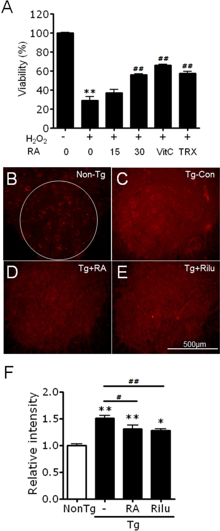
Fig. 3. Rosmarinic acid treatment suppresses oxidative stress in vitro and in the lumbar cords of G93A-SOD1 transgenic mice. (A) Rosmarinic acid treatment suppressed H2O2 (800 µM)-induced cytotoxicity in SH-SY5Y neuroblastoma cells. Cells were treated with 15 or 30 µg/ml rosmarinic acid (RA). Vitamin C (VitC) and trolox (TRX) were used at 200 µM. Cell viability was measured using the WST-1 assay after 24 h of treatment. Data points represent means±SEM (n=6). **denotes difference compared to untreated control at p<0.01. ##denotes difference compared to H2O2-treated cells at p<0.01. One-way ANOVA and Newman-Keuls post hoc test were used. (B~F) Photomicrographs showing anti-HNE-stained ventral horns of the lumbar cord of non-transgenic control mice (Non-Tg; B), G93A-SOD1 transgenic control mice (Tg-Con; C), G93A-SOD1 transgenic mice treated with 400 mg/kg/day rosmarinic acid (Tg+RA; D), and G93A-SOD1 mice treated with 35 mg/kg/day riluzole (Tg+Rilu; E). (F) Fluorescence levels in the ventral horns [circled area in (B), 600 µm in diameter] of the lumbar cord of each group were quantified. All animals were examined at 16 weeks of age. Scale bar; 500 µm. Data are presented as means±SEM (n=8~13). * and ** denote differences compared to non-transgenic control (NonTg) at p<0.05 and p<0.01, respectively. # and ## denote difference compared to Tg-CON at p<0.05 and p<0.01, respectively. One-way ANOVA and Newman-Keuls post hoc test were used.
© Exp Neurobiol