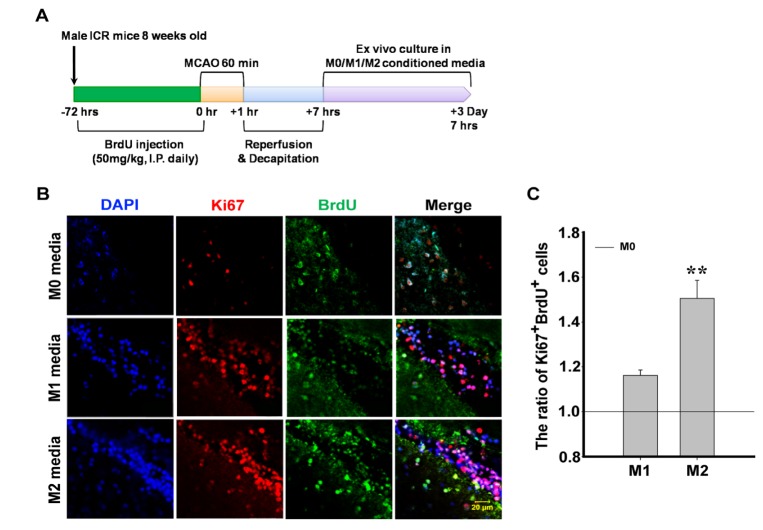
Fig. 2. Proliferation SVZ-NSPCs in M2 conditioned media. (A) Schematic time course of the in vivo and ex vivo experiments. (B) Immunohistochemistry of Ki67 and BrdU in M0, M1, and M2 conditioned media shows the proliferation of SVZ-NSPCs in THE organotypic brain. (C) Quantification graph of the ratio of Ki67+ and BrdU+ cells of SVZ-NSPCs in organotypic brain (Scale bar=20 um, **p<0.05 vs. M0 conditioned media, n=5/group).
© Exp Neurobiol