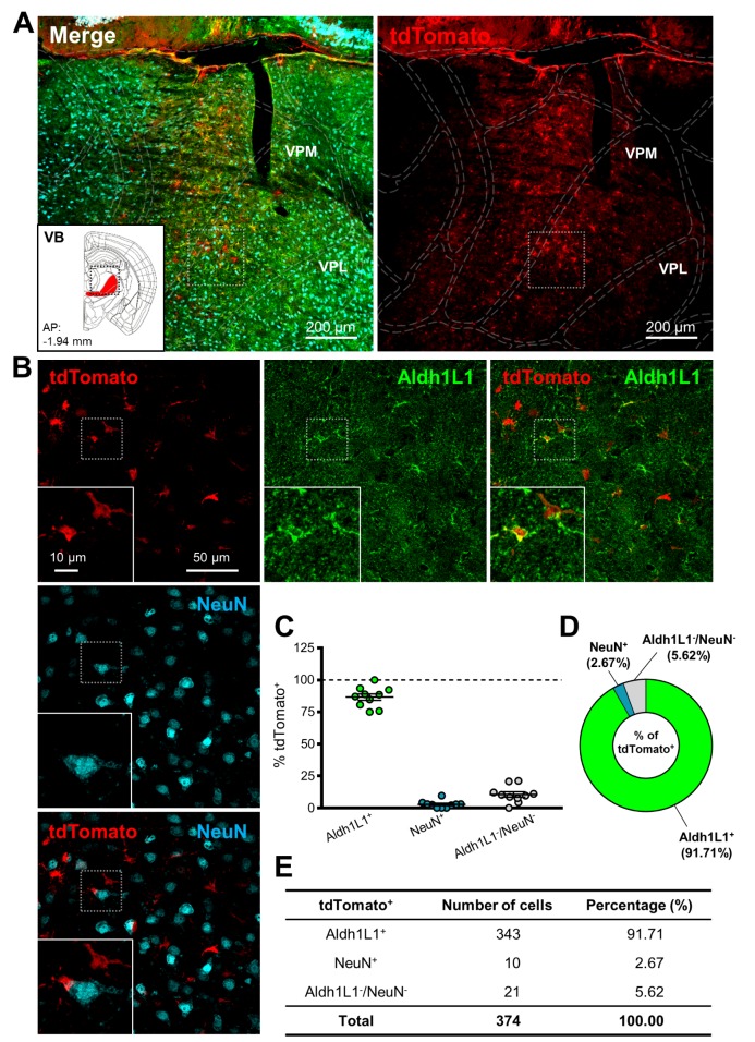
Fig. 2. hALDH1L1 promoter-driven Cre recombinase-mediated tdTomato expression in thalamic VB. (A) Low magnification images of injection area in thalamus. Each color represents as following description. Green indicates immunoreactivity of Aldh1L1 protein; Red indicates tdTomato expression; Cyan indicates immunoreactivity of NeuN. Inset indicates the area of target (red area, VB). (VPM, ventral posteromedial nucleus; VPL, ventral posterolateral nucleus). (B) High magnification images from the box in (A). (C) Summary graph of percentages of Aldh1L1+ (green) cells and NeuN+ (cyan) cells (and also both negative cells, gray) in tdTomato+ cells from each image (Mean±SEM). (D) Quantification of tdTomato+ population with Aldh1L1+, NeuN+ and Aldh1L1-/NeuN-cells in thalamic VB area. (E) Summary of tdTomato+ cells in thalamic VB area.
© Exp Neurobiol