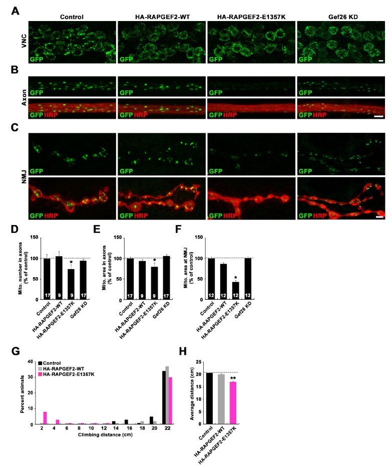
Fig. 5. Overexpression of the human RAPGEF2-E1357K variant in Drosophila motor neurons disrupts the distribution of mitochondria to axons and NMJ terminals. (A~C) Confocal images of ventral nerve cord (VNC, A), axon (B), and NMJ 6/7 (C) in third instar larvae stained with anti-GFP alone (A) or together with anti-HRP (B and C). Axons and NMJs in abdominal segment 5 were analyzed. Genotypes include D42-GAL4,UAS-mito-GFP/+ (control), D42-GAL4,UAS-mito-GFP/UAS-HA-RAPGEF2-WT(HA-RAPGEF2-WT), UAS-HA-RAPGEF2-E1357K/+; D42-GAL4,UAS-mito-GFP/+ (HA-RAPGEF2-E1357K), and D42-GAL4,UAS-mito-GFP/UAS-gef26RNAi(Gef26 KD). Scale bars, 5 µm. (D~F) Quantification of the number (D) and area (E and F) of Mito-GFP-positive puncta in axons (D and E) and at NMJ terminals (F). Values were normalized to the respective area of axon or NMJ terminals. (G) Distribution of the distance climbed by 20-day-old D42-GAL4/+ (control), D42-GAL4/UAS-HA-RAPGEF2-WT (HA-RAPGEF2-WT), and UAS-HA-RAPGEF2-E1357K/+; D42-GAL4/+ (HA-RAPGEF2-E1357K) flies over 30 s. (H) Quantification of average climbing distance. Data are presented as mean±SEM. All comparisons are made with the D42-GAL4,UAS-mito-GFP/+ control (*p<0.001; **p<0.01).
© Exp Neurobiol