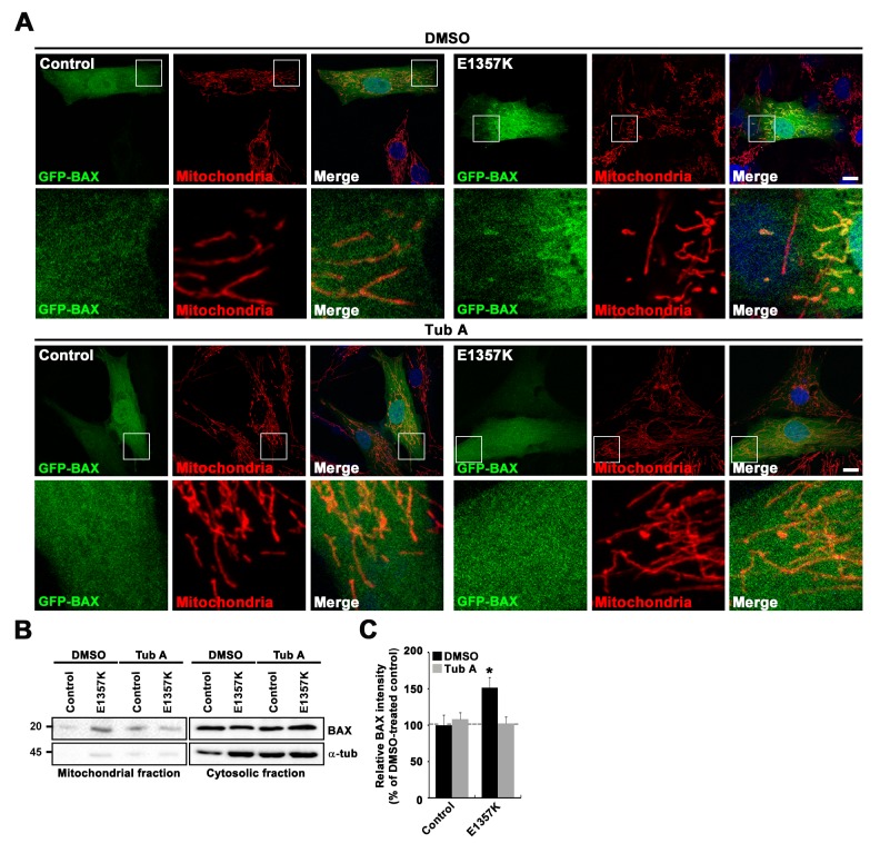
Fig. 7. Pharmacological inhibition of HDAC6 restores abnormal accumulation of BAX in the mitochondria in ALS patient-derived fibroblasts. (A) Confocal images of control and patient (E1357K) fibroblasts expressing GFP-BAX stained with anti-mitochondria antibody and DAPI. Cells were treated with DMSO or 1 µM tubastatin A (Tub A) prior to immunostaining. Scale bar, 5 µm. (B) Western blot analysis of mitochondrial and cytosolic fractions from DMSO- or Tub A-treated control and patient fibroblasts using anti-BAX and anti-α-tubulin antibodies. (C) Quantitative analysis of densitometric measurements (n=3). The band intensities of BAX in each fraction were normalized to the sum of mitochondrial and cytosolic BAX intensities. Data are presented as mean±SEM. Comparisons are made with the DMSO-treated control (*p<0.001).
© Exp Neurobiol