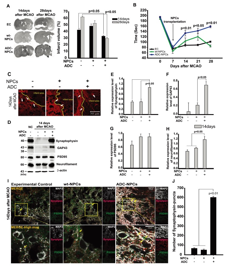
Fig. 6. Transplanted neural progenitor cells (NPCs) overexpressing arginine decarboxylase (ADC) genes (ADC-NPCs) attenuate infarct volume and promote motor function following middle cerebral artery occlusion (MCAO). (A) Infarct volume analysis by hematoxylin and eosin (H&E) staining. The graph summarizes the significant changes in the transplanted ADC-NPCs following cerebral ischemia. (B) Rotarod test conducted 7~28 days after MCAO. The graph represents neurological motor function recovery following cerebral ischemia (n=12 per condition). (C) Immunocytochemical staining of MAP2 protein in the ipsilateral striatum 14 days after MCAO. (D) Immunoblotting of the neural differentiation- and synapse formation-related proteins in the ipsilateral striatum 14 days after MCAO. (E–H) Quantification graphs of immunoblotting summarizing the expression levels of synapse formation-related proteins, normalized to the control. (I) High-magnification image of the results of immunocytochemical staining showing MAP2 (white)/synaptophysin (red)/PSD95 (green)-positive cells in the ipsilateral striatum 14 days after MCAO. (J) Quantification graph of the number of synaptophysin puncta in the ipsilateral striatum 14 days after MCAO. EC, experimental control; NPCs, transplanted neural stem cells; ADC-NPCs, neural progenitor cells (NPCs) overexpressing arginine decarboxylase (ADC) genes. Yellow arrows indicate colocalized synaptophysin puncta and PSD95 in the MAP2-positive cells. The error bars represent the mean±SEM.
© Exp Neurobiol