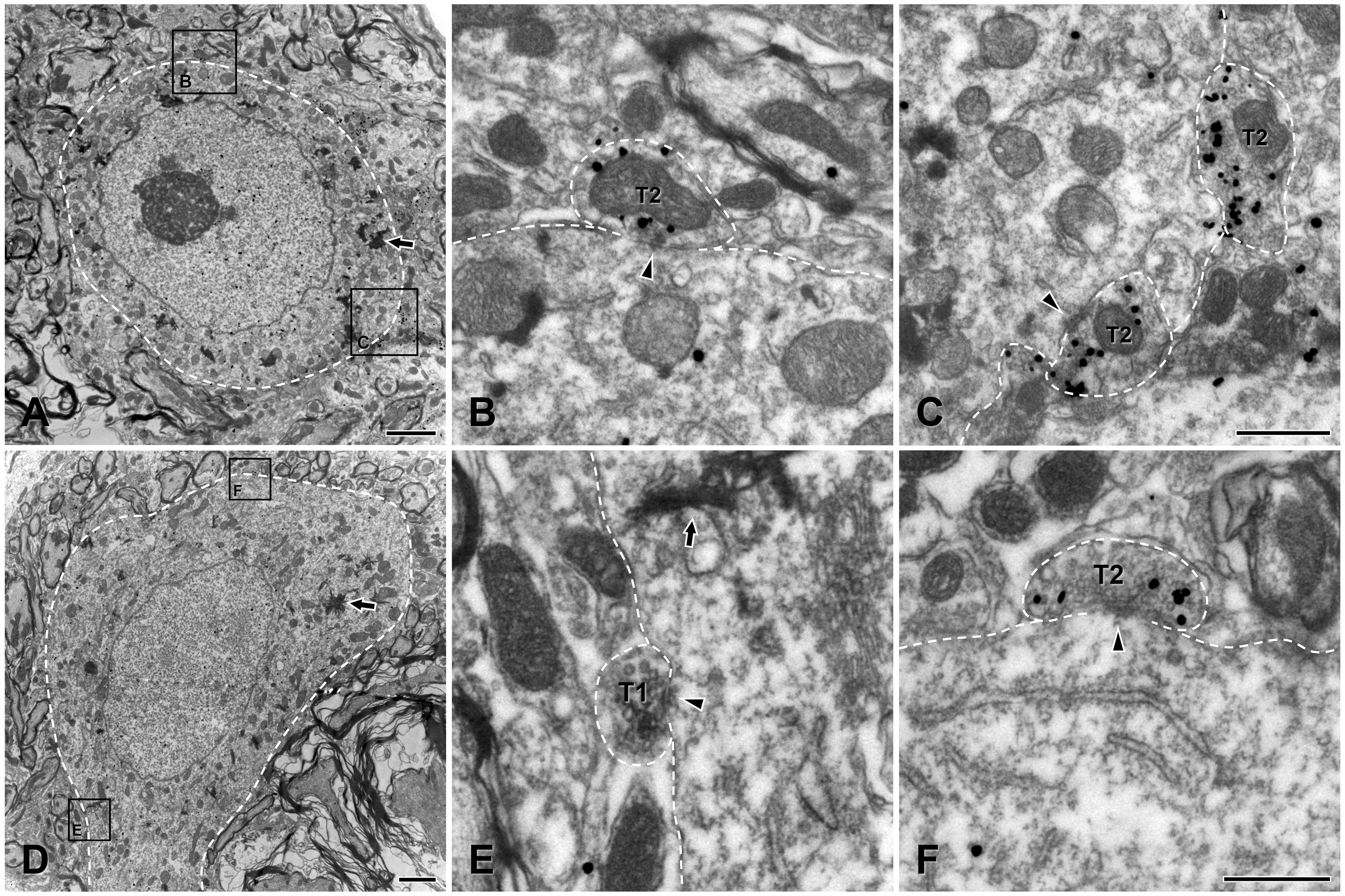
Fig. 2. Electron micrographs showing two (A~C and D~F) HRP-labeled, jaw-closing γ-motoneurons in the trigeminal motor nucleus (the reaction product of retrogradely-transported HRP is indicated by arrows), contacted by VGLUT1+ bouton (T1, peroxidase labeling) and VGLUT2+ boutons (T2, gold-silver labeling). B and C are enlargements of the boxed areas in A; E and F are enlargements of the boxed areas in D. The boundaries of the somata of the motoneurons and the VGLUT+ boutons in contact with them are outlined with a dashed line. Arrowheads indicate synapses. Scale bars, 2 μm in A and D, and 500 μm in B, C, E and F.
© Exp Neurobiol