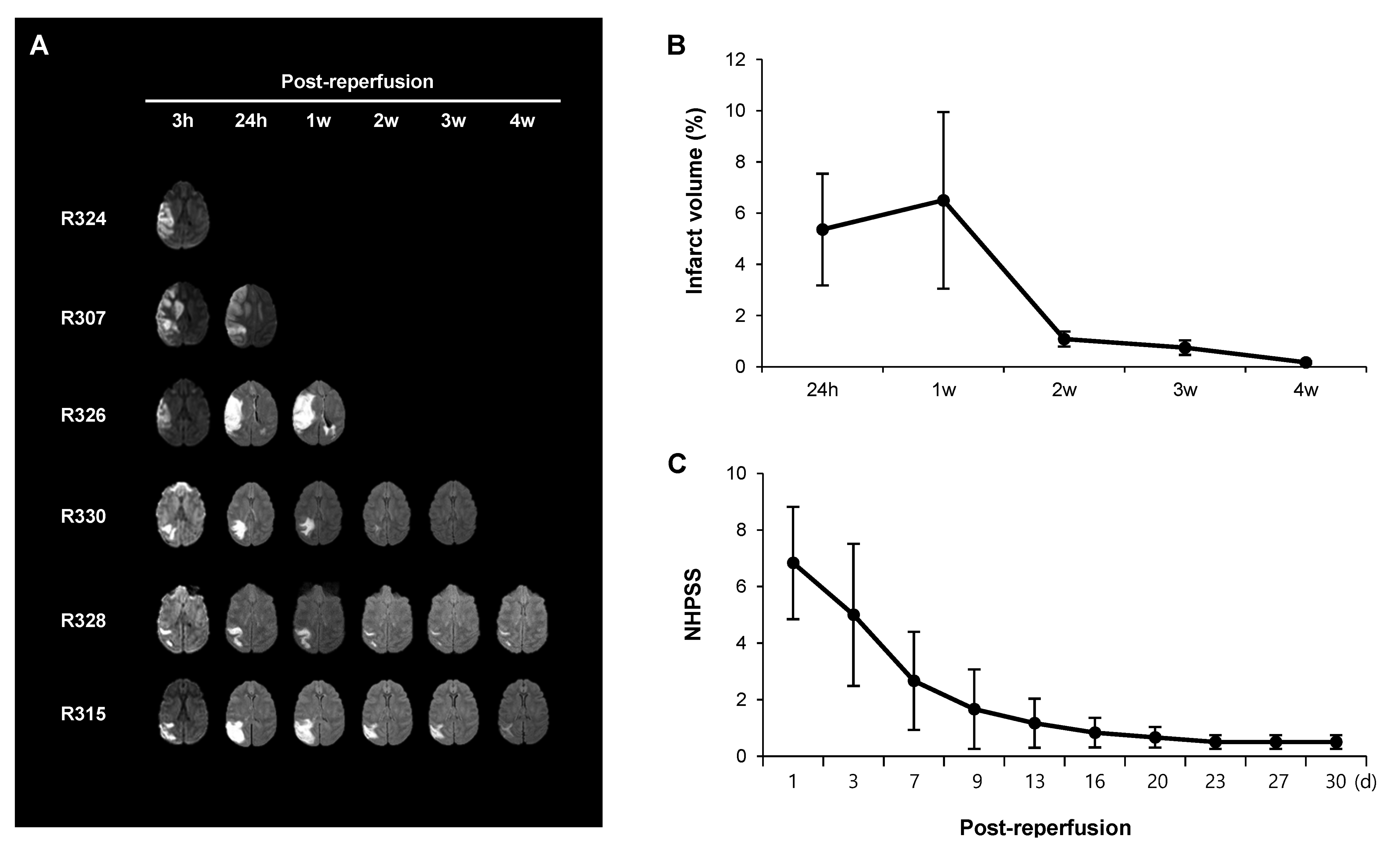
Fig. 2. Gradual reduction in the infarct volume is associated with neurological recovery. (A) Follow-up MRI with fluid-attenuated inversion recovery (FLAIR) imaging was performed in six rhesus monkeys from 24 hours until 4 weeks after reperfusion. Diffusion weighted imaging is shown at 3 hours after reperfusion. (B) Changes in the infarct volume calculated from the FLAIR images in the subacute and chronic groups (R326, R330, R328, and R315). (C) Changes in the neurological scores measured by Non-human Primate Stroke Scale (NHPSS) in the chronic group (n=5). Data are presented as mean±standard error of mean (SEM).
© Exp Neurobiol