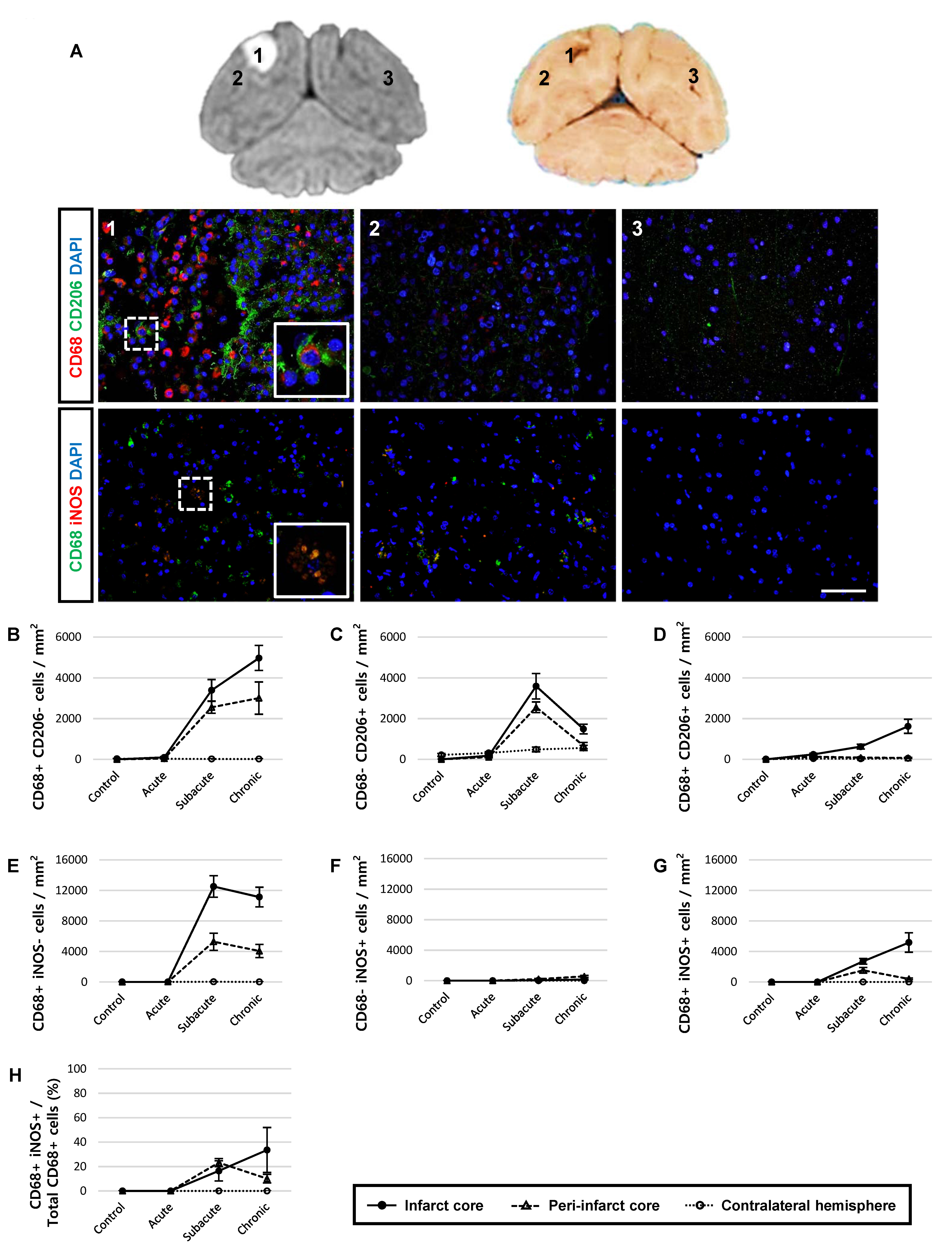
Fig. 6. CD68-positive phagocytic cells have low correlation with the M2 type microglia/macrophage marker, CD206. (A) Paraffin-embedded brain sections were immunostained with CD68 for phagocytic cells, CD206 for M2 type, and iNOS for M1-type microglia/macrophages, followed by coun-terstaining with DAPI for cell nuclei at one month after reperfusion. Merged cells (inset image) were specifically detected in the subacute and chronic stage. 1, infarct core; 2, peri-infarct core; 3, contralateral hemisphere. Representative images were obtained from animal R330. Scale bar represents 10 μm. (B~D) Quantification of CD68- and CD206-positive cells. The number of CD68-specific (B), CD206-specific (C), and CD68+/CD206+-coexpressing (D) cells was quantified using Image J cell counter. (E~G) Quantification of CD68- and iNOS-positive cells. CD68-specific (E), iNOS-specific (F), and CD68+/iNOS+-co-expressing (G) cells were detected. (H) The ratio of CD68+/iNOS+ cells over the total CD68+ cells are shown. Data are presented as mean±SEM. Each line chart symbol represents 50 regions of interest, with 2 animals in each group.
© Exp Neurobiol


