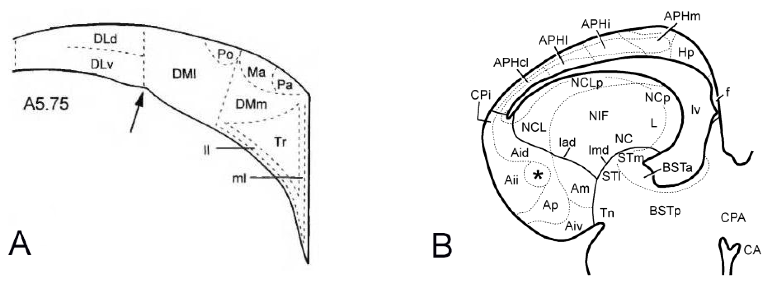
Schematic illustration of subdivisions respectively in the pigeon hippocampal formation (A) and chicken embryo brain (B) [2, 11].