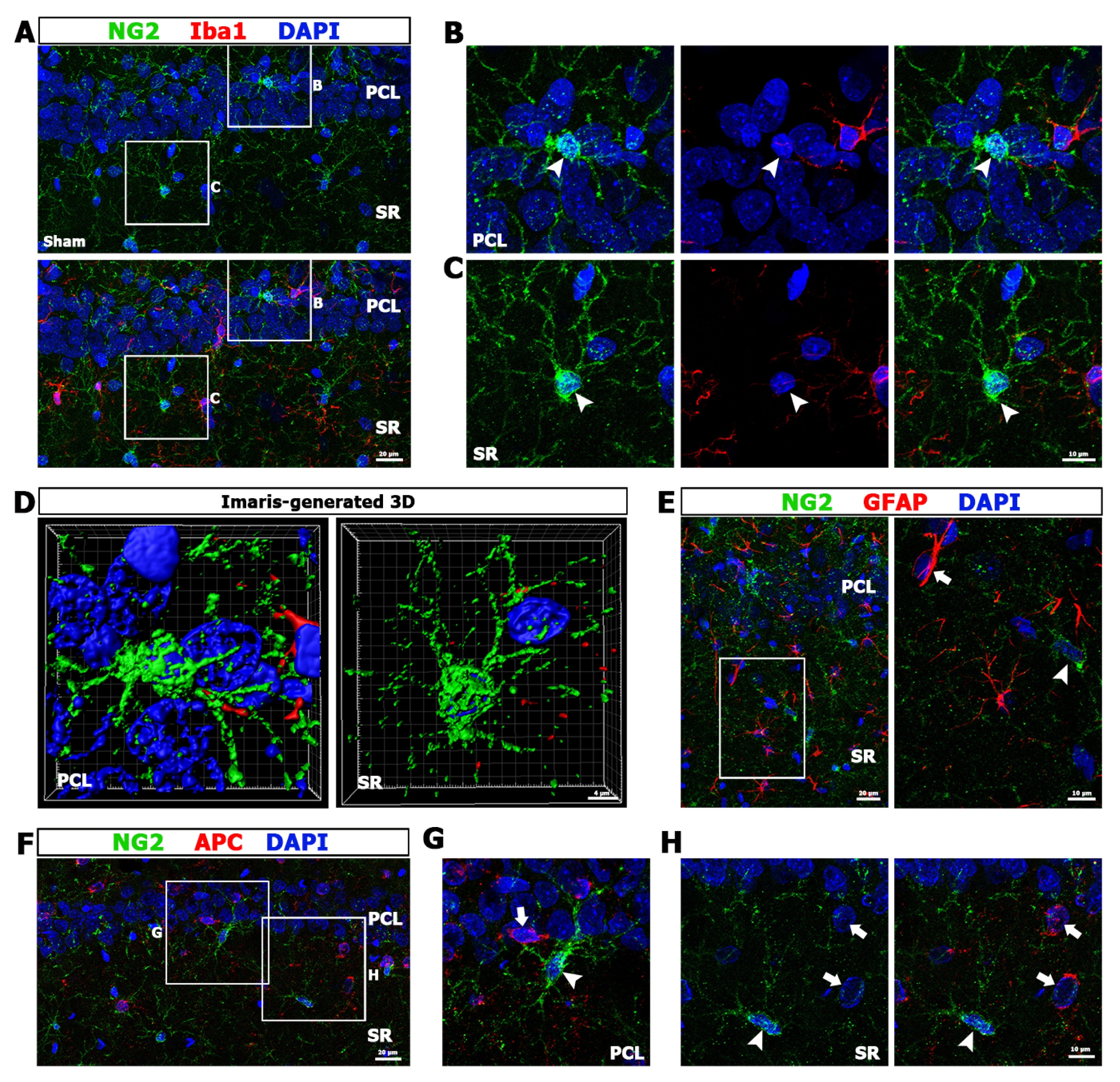
Characterization of NG2 glia in the hippocampal CA1 region of sham-operated rats using double-labeling for NG2 and Iba1. (A~C) Lower- (A) and higher- (B, C) magnification views. The boxed areas of the pyramidal cell layer (PCL) and stratum radiatum (SR) in A are enlarged in B and C, respectively. NG2 glia and Iba1-positive microglia do not have overlapping distributions in the CA1 hippocampus. Note that resting NG2 glia in the pyramidal cell layer (arrowheads in B) and the stratum radiatum (arrowheads in C) have multiple fine processes radiating in all directions. (D) Threedimensional renderings of the images shown in B and C, showing that NG2 glia share similar morphological characteristics in both strata of the CA1 hippocampus. (E) Double-labeling for NG2 and GFAP in the CA1 hippocampus of sham-operated rats. The boxed area in the left panel is enlarged in the right panel. Note that NG2 glia (arrowhead) and astrocytes (arrow) do not have overlapping distributions in the CA1 hippocampus. (F~H) Doublelabeling for NG2 and APC, a marker of mature oligodendrocytes, in the CA1 hippocampus of sham-operated rats. The boxed areas of the pyramidal cell layer and stratum radiatum in F are enlarged in G and H, respectively. Note that NG2 glia (arrowheads) are distinct from mature oligodendrocytes (arrows). Cell nuclei are stained with DAPI. Scale bars represent 20 μm in A, left panel of E and F; 10 μm in B, C, right panel of E, G and H; and 4 μm in D.