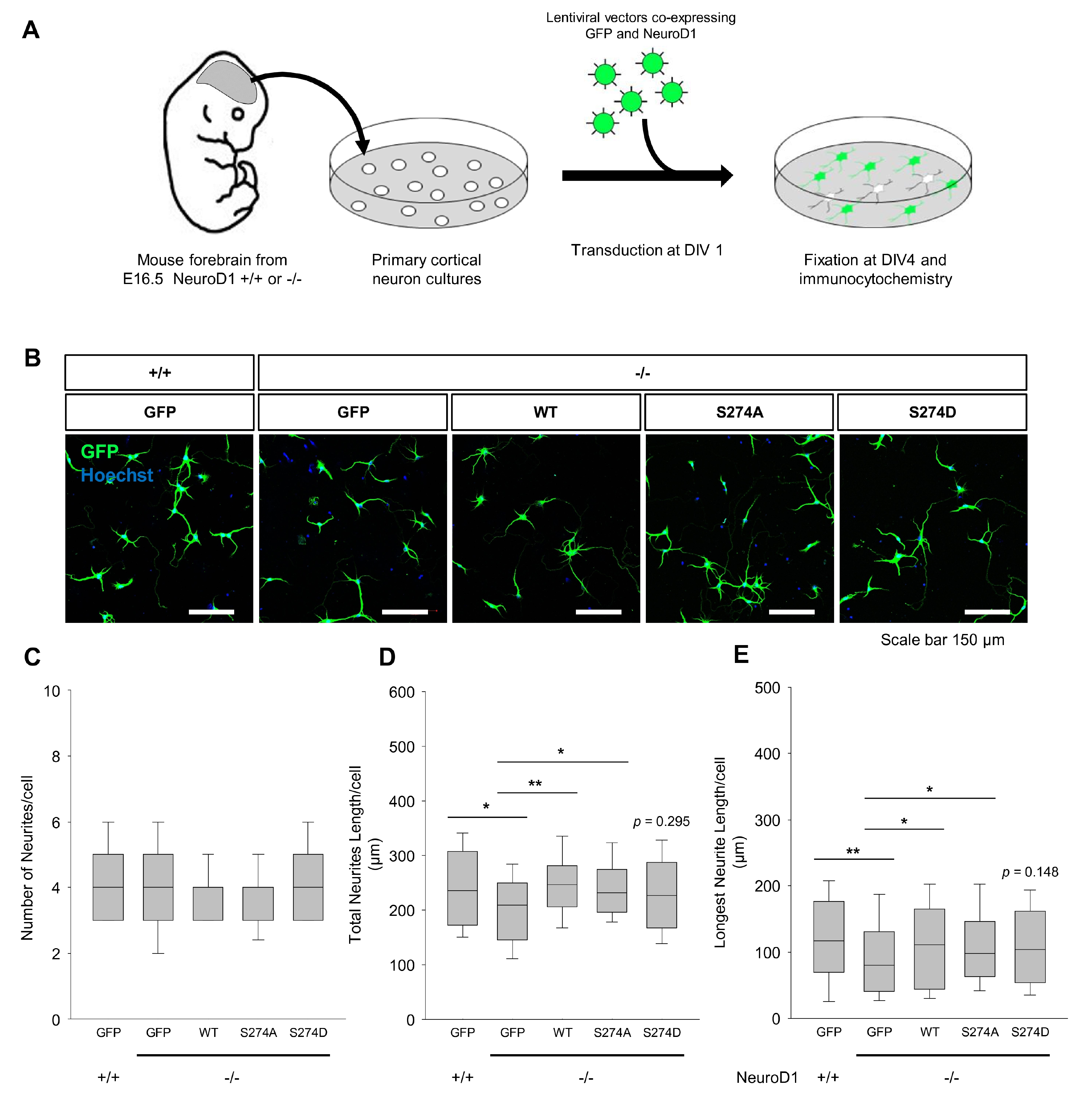
Fig. 6. Rescue experiments to supplement neurogenic differentiation 1 (NeuroD1) deficiency. (A) Schematic representation of the experimental process for transducing primary cortical neurons from the brain of NeuroD1 knockout (KO) embryos. One day after plating, cortical neurons were transduced with lentiviral vectors encoding green fluorescent protein (GFP) together with wild type (WT) or S274 mutants. Cells were fixed at the 5th day in vitro (DIV). (B) Cells were immunostained with anti-GFP antibody (green) to detect lentivirus transduced neurons and Hoechst (blue) for nuclei. (C~E) Neurite length was quantified from images shown in (B) using the NeuronJ plugin of the ImageJ software program. The number of neurites, total neurite length, and longest neurite length per cell were counted and presented. Data are represented as means±S.E.; at least 50 neurons were counted per group (*p<0.05; **p<0.01, compared to the value of KO neurons expressing GFP as a control).
© Exp Neurobiol