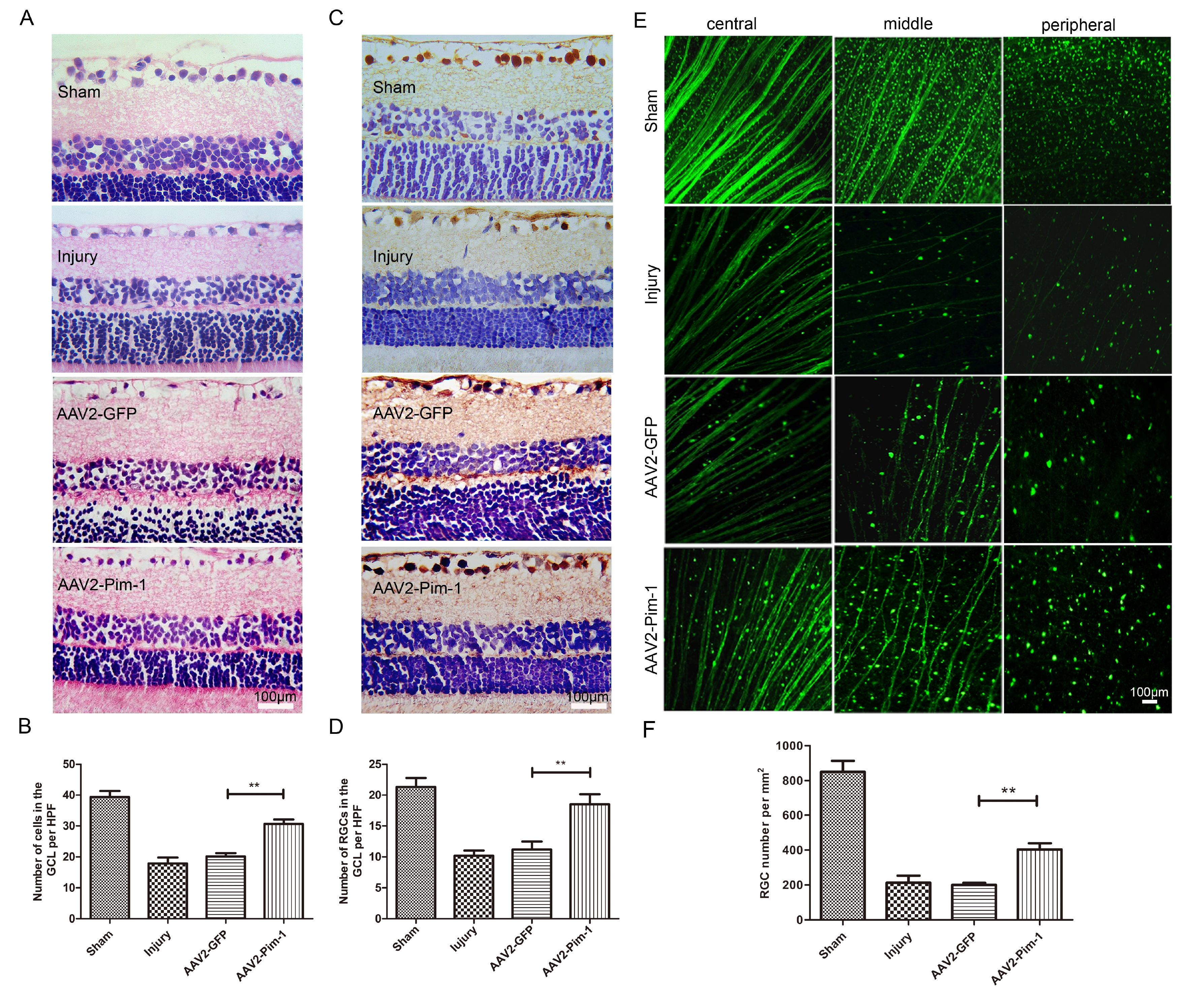
Fig. 3. HE staining, γ-synuclein immunohistochemical staining and FITC-CTB tracing to test survival of RGCs. (A, C and E) All staining of retinal sections 2 weeks after ONC, and γ-synuclein-positive RGCs were visualized in brown yellow, RGC density in the representative flat-mounted retina by FITC-CTB tracing to test survival of RGCs. (B, D and F) AAV2-mediated overexpression of Pim-1 showed a significant neuroprotective effect on RGCs. Compared with AAV2-GFP group, **p<0.01; n=6.
© Exp Neurobiol