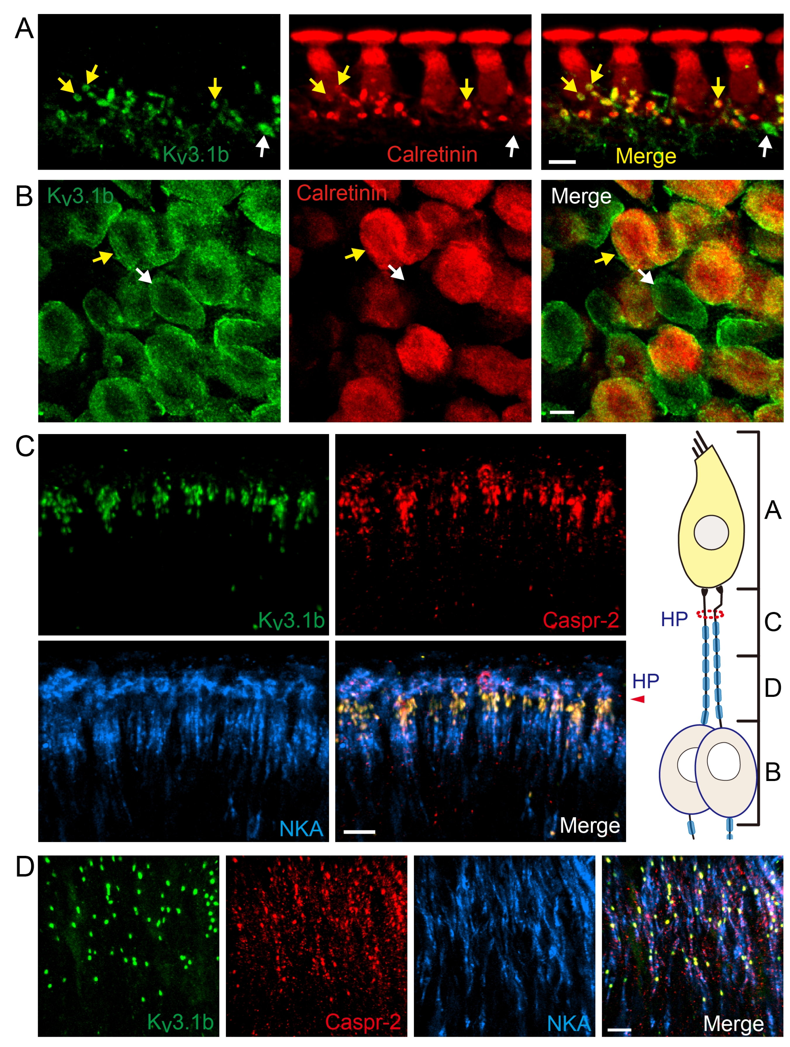
Fig. 2. Kv3.1b is expressed in the unmyelinated dendritic segments and the nodes of Ranvier of the type 1 cochlear afferent nerve fibers. The relative anatomical regions corresponding to each panel A-D are described in the diagram. Scale bar: 10 μm. (A) IHC-type 1 afferent fiber synapses immunolabeled with anti-Kv3.1b and anti-calretinin (n=6 cochlear preparations from 4 rats). Kv3.1b signal is found at the cochlear afferent fibers’ dendritic terminals (yellow arrow) where they contact the IHCs. Kv3.1b-postive but calretinin-negative dendritic terminal (white arrow) is also found. (B) Spiral ganglion neurons (SGNs) immunolabeled with anti-Kv3.1b and anti-calretinin (n=4 cochlear preparations from 4 rats). Kv3.1b signal is found in both calretinin-positive (yellow arrow) and calretinin-negative (white arrow) neurons. Calretinin signal is present in 62.2% of Kv3.1b-positive SGNs (305 out of 490 Kv3.1b-positive SGNs). (C, D) Type 1 afferents immunolabeled with anti-Kv3.1b, anti-Caspr-2, and anti-NKA (n=6 cochlear preparations from 3 rats). Kv3.1b-hot spots along the nerve fiber correspond well with Caspr-2-signal.
© Exp Neurobiol