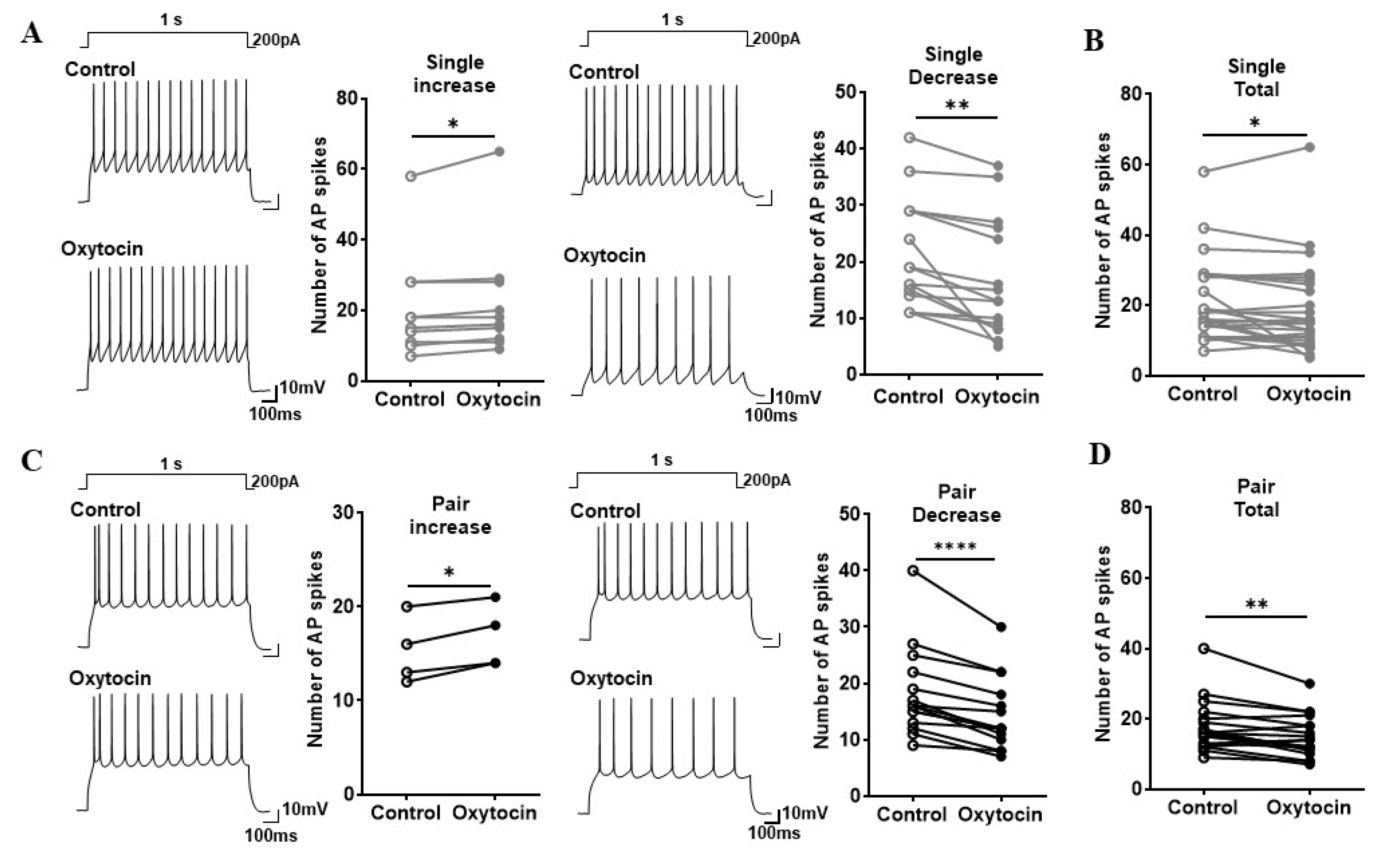
Fig. 3. Similar changes in the pattern of oxytocin-induced AP spike number in pyramidal neurons in single- and pair-exposed rats. (A, B) Single-exposed rats. (C, D) Pair-exposed rats. (A, C) Representative AP spike traces in pyramidal neuron to a depolarizing pulse (200 pA current injection for 1s) in the absence (Control) and the presence of oxytocin (Oxytocin), and a graph of the statistical values (Left, increase; Right, decrease) are shown (Paired t-test, *p<0.05, **p<0.01; ****p<0.0001). AP, action potential. (B, D) Total number of AP spikes (Paired t-test, *p<0.05, **p<0.01).
© Exp Neurobiol