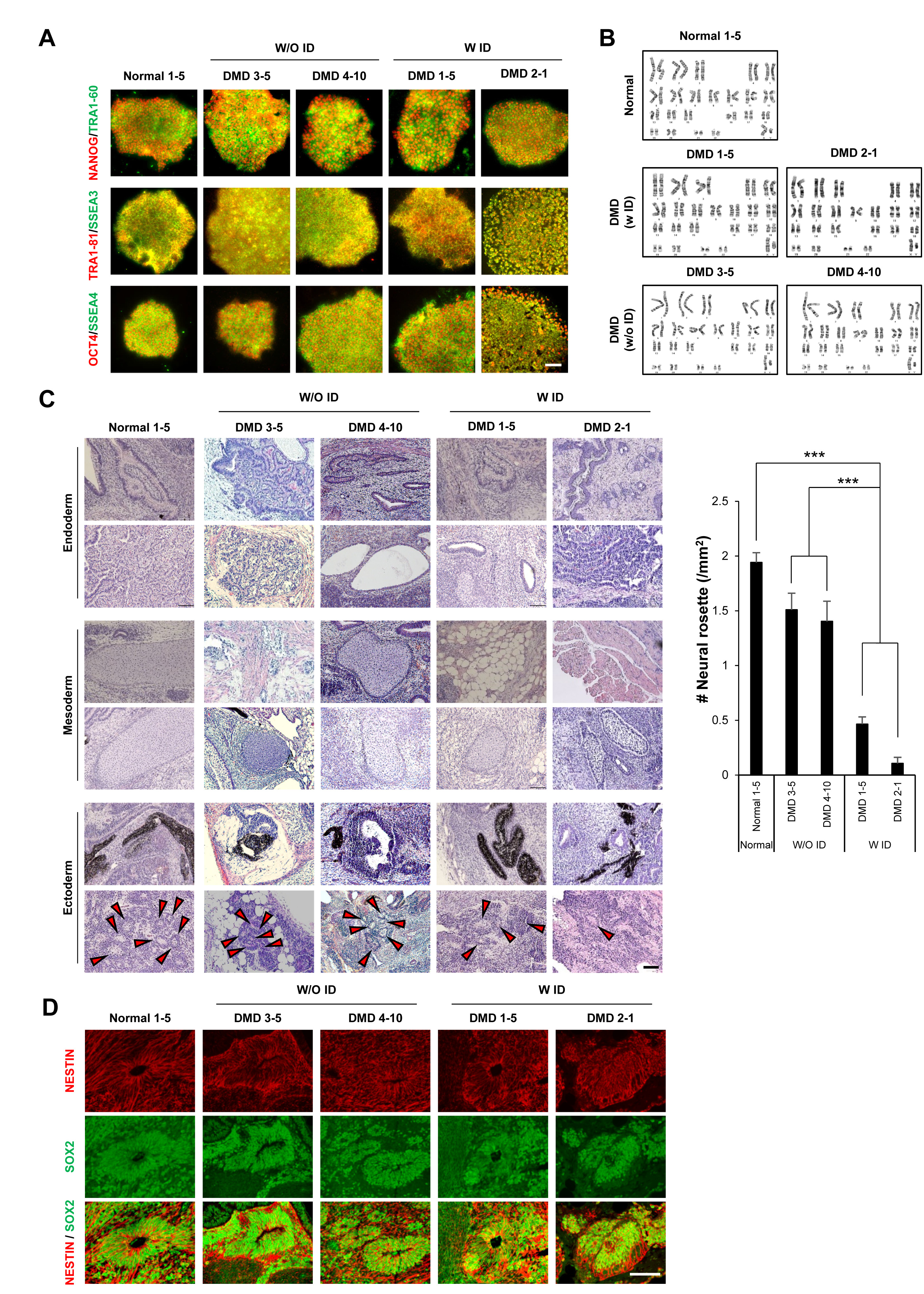
Fig. 1. iPSCs derived from DMD patients with ID show defects in neural ectoderm formation. (A) Immunofluorescence staining shows that stem cell markers (OCT4, NANOG, SSEA3/4, and TRA 1-60/1-81) are well expressed in the iPSCs generated from all five individuals. (B) Karyotyping analysis data show normal karyotypes in iPSCs derived from all five participants. (C) Hematoxylin and eosin staining of teratomas shows that iPSCs from all individuals were able to differentiate into the three germ layer types (endoderm, mesoderm, and ectoderm). However, fewer neural rosettes (arrowheads) were formed by cells derived from DMD patients with ID (W/ID) than from those from a normal individual or DMD patients without ID (W/O ID). (D) Immunofluorescence staining of teratomas shows neural rosettes in teratomas successfully expressed NESTIN and SOX2. Scale bars: (A) 50 μm, (C, D) 100 μm. Values denote means±SEMs. One-way ANOVA with Bonferroni’s post hoc test. ***p<0.0001.
© Exp Neurobiol