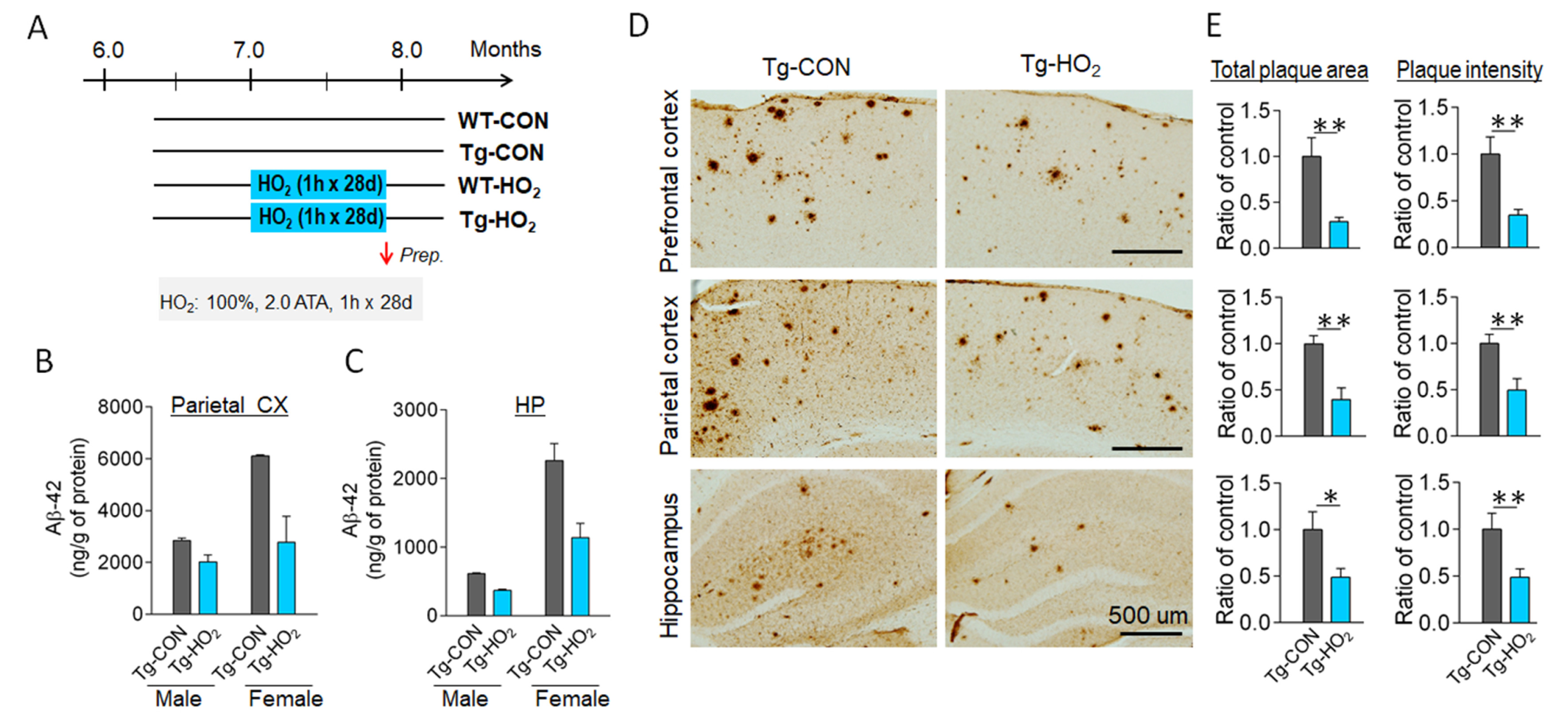
Fig. 1. Hyperoxygenation treatment suppressed beta-amyloid plaque deposition in the brain of APP/PS1-Tg mice. (A) Experimental design. Tg-APP/PS1 mice at 7 months of age were exposed to 100% O2 at 2 ATA for 60 min daily for 28 days. (B, C) ELISA data showing insoluble Aβ(1-42) levels in the parietal cortex (B) and hippocampus (C) of Tg-APP/PS1 control mice (Tg-CON) and Tg-APP/PS1 mice exposed to hyperoxygenation (Tg-HO2). Male and female mice were separately examined. n=4~7 animals/group, 6~8 repeats. (D, E) Immunohistochemical images showing plaque deposition stained with Bam-10 anti-Aβ antibody in the prefrontal cortex, parietal cortex, and hippocampus of Tg-CON and Tg-HO2 (D). Quantification levels by total plaque areas (relative ratios of summed plaque area in μm2/mm2) and summed stained intensity (relative ratios of arbitral unit) were presented (E). n=5~7 animals/group, 10~12 sections. Data are presented as mean±SEM. *,**difference between indicated groups; *p<0.05; **p<0.01 (Student’s t-test).
© Exp Neurobiol