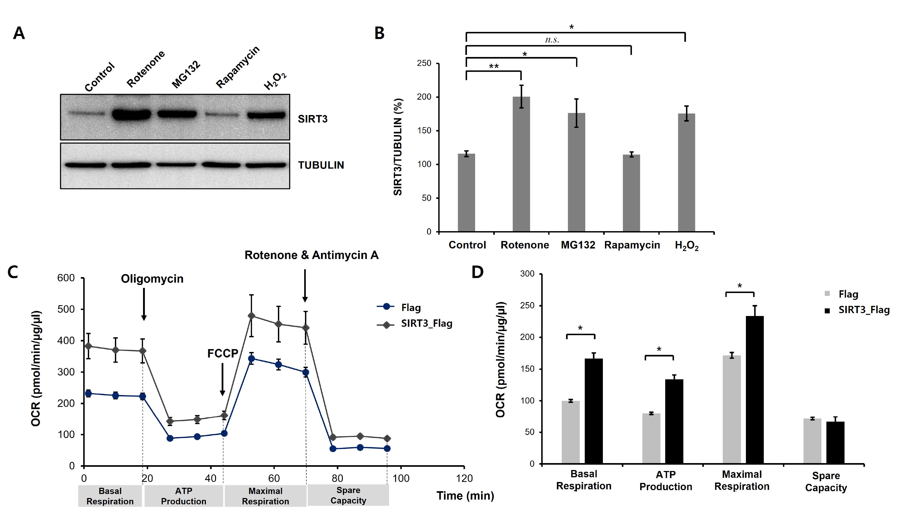
Fig. 1. Neurotoxic stress increases SIRT3 protein levels in differentiated SH-SY5Y cells. (A) SH-SY5Y cells were treated with all-trans retinoic acid (RA, 10 µM) for 8 days to generate differentiated SH-SY5Y cells. Western blot analysis was performed to measure SIRT3 protein expression. Differentiated SH-SY5Y cells were treated with rotenone (50 µM), MG132 (5 µM), rapamycin (10 µM), and H2O2 (500 µM) for 24 h, and then the cells were harvested for total protein extraction. Rotenone, MG132, and H2O2 significantly increased SIRT3 levels in differentiated SH-SY5Y cells. (B) Quantification of the immunoblots was performed from 3 independent experiments. Tubulin was used for normalization. *p<0.05; **p<0.005; n.s., not significant (one-way ANOVA with Tukey’s post-hoc comparison test). (C) Stable Flag- or human SIRT3-Flag-expressing cell lines were treated with RA (10 µM) for 8 days, and then mitochondrial oxygen consumption was analyzed. Mitochondrial dysfunction of Flag- or SIRT3-Flag-expressing differentiated SH-SY5Y cells was measured by detecting the basal OCR, ATP production, maximum reserve, and respiratory capacity by a Seahorse XF Analyzer. The OCR was normalized to the total protein concentration. (D) Basal respiration, ATP production, maximum respiration, and spare respiratory capacity were quantified and are shown as percentages of the basal values. Data are presented as the mean±SEM. *p<0.05 (unpaired Student’s t-test).
© Exp Neurobiol