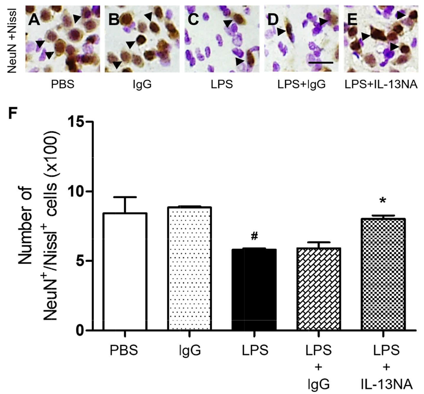
Fig. 1. Interleukin-13 contributes to neurodegeneration of LPS-injected rat striatum in vivo. Animals unilaterally received intrastriatal injection of PBS (A, 3 µl) or non-specific IgG (IgG) only (B, 1 µg) as a control, LPS (C, 5 µg), LPS+IgG (D, 1 µg) and LPS+Interleukin 13 neutralizing antibody (IL-13NA) (E, 1 µg). (A~E) At 7 days after LPS injection, animals were sacrificed, and the coronal sections (40 µm) were selected and processed for neuronal nuclei (NeuN) immunohistochemical staining and Nissl staining. Arrowheads indicate NeuN+ cells merged with Nissl+ cells (NeuN+/Nissl+). (F) Number of NeuN+/Nissl+ cells in the striatum at 7 days after LPS injection. #p<0.001, as compared with PBS, *p<0.001, as compared with LPS+IgG. One-way ANOVA and Newman–Keuls analyses. Four to eleven animals were used for each experimental group. The results represent mean±SEM. Scale bar, (A~E) 25 µm.
© Exp Neurobiol