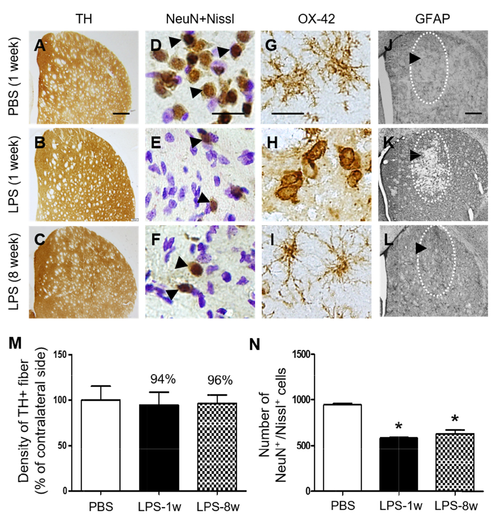
Fig. 7. Temporal effects of LPS on dopamine fibers and inflammation in the striatum in vivo. Animals unilaterally received intrastriatal injection of PBS (A, D, G, J, 3 μl) as a control and LPS (B, C, E, F, H, I, K, L, 5 μg/3 μl). (A~C) At 1 week after LPS injection, animals were sacrificed and the coronal sections (40 μm) were selected and processed for tyrosine hydroxylase (TH) immunohistochemical staining, (D~F) neuronal nuclei (NeuN) immunohistochemical staining and Nissl staining, (G~I) OX-42 immunohistochemical staining (insert images are ED1 Immunohistochemical staining) and (J~L) GFAP immunohistochemical staining. (D~F) Arrowheads indicate NeuN+ cells merged with Nissl+ cells (NeuN+/Nissl+). (J~L) Dotted round indicates area lacking in GFAP+ astrocytes. Arrow indicates syringe track. (M) Density of TH+ fibers in the striatum at 1 week after intrastriatal injection of PBS as a control (A) or at 1 week (B) and 8 weeks (C) after intrastriatal injection of LPS. Density of striatal TH+ fibers was not significantly affected by LPS in rat striatum. One-way ANOVA and Newman–Keuls analyses. Three animals were used for each experimental group. The results represent mean±SEM. (N) Number of NeuN+/Nissl+ cells in the striatum at 1 week after intrastriatal injection of PBS as a control (D) or at 1 week (E) and 8 weeks (F) after intrastriatal injection of LPS. *p<0.001, as compared with PBS. One-way ANOVA and Newman–Keuls analyses. Four to five animals were used for each experimental group. The results represent mean±SEM. Scale bar, (A~C) 500 µm; (D~I) 20 µm; (J~L) 500 µm.
© Exp Neurobiol