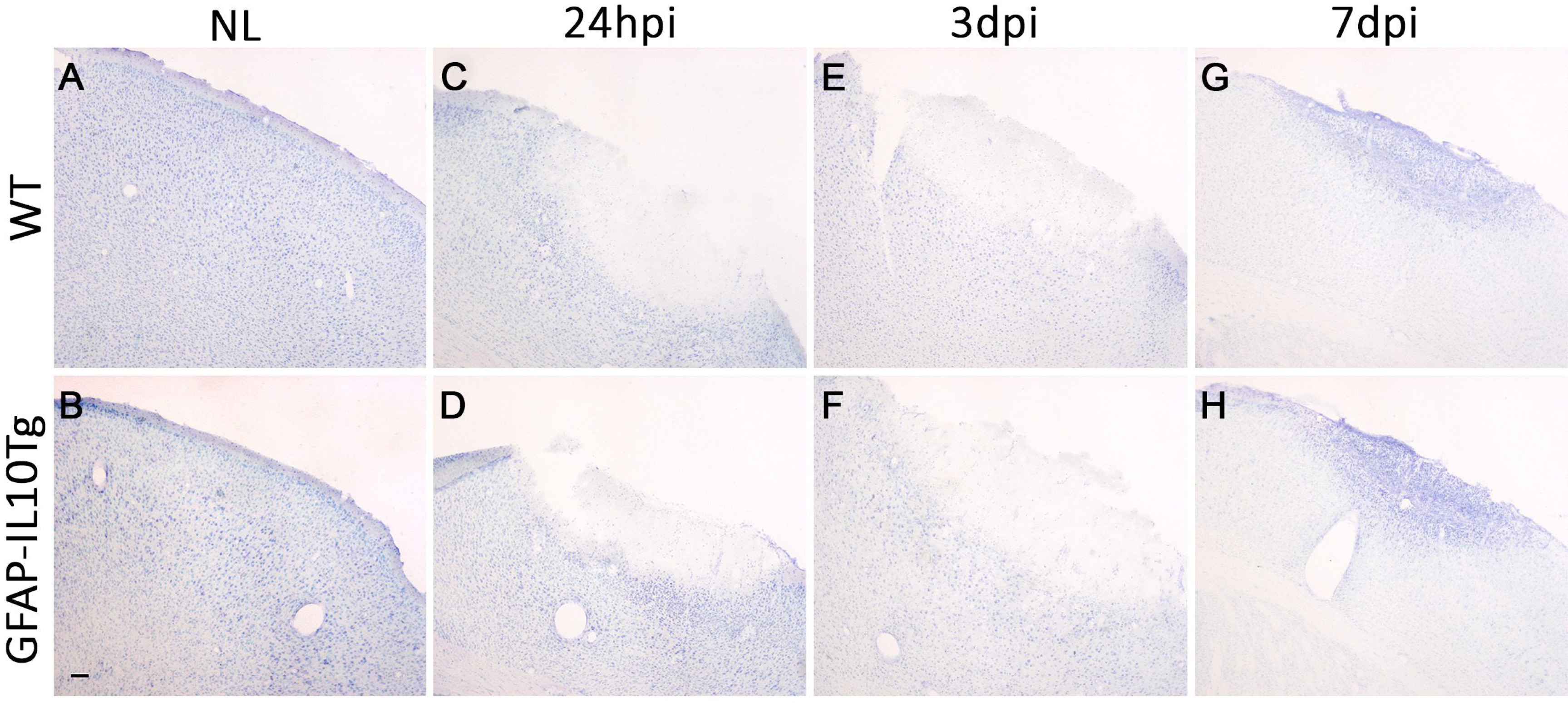
Fig. 2. Toluidine blue staining. Representative images showing the toluidine blue staining of cortex in non-lesioned (NL) WT (A) and NL GFAP-IL10Tg (B) animals. After TBI, both WT (C, E, G) and GFAP-IL10Tg animals (D, F, H) showed the area of lesion in the cortex characterized by a depleted area of neurons surrounded by the penumbra, exhibiting pigmented nuclei indicative of degeneration from 24 hpi to 7 dpi. No differences between WT and GFAP-IL10Tg animals were detected at any time-point studied. Scale bar=100 µm.
© Exp Neurobiol