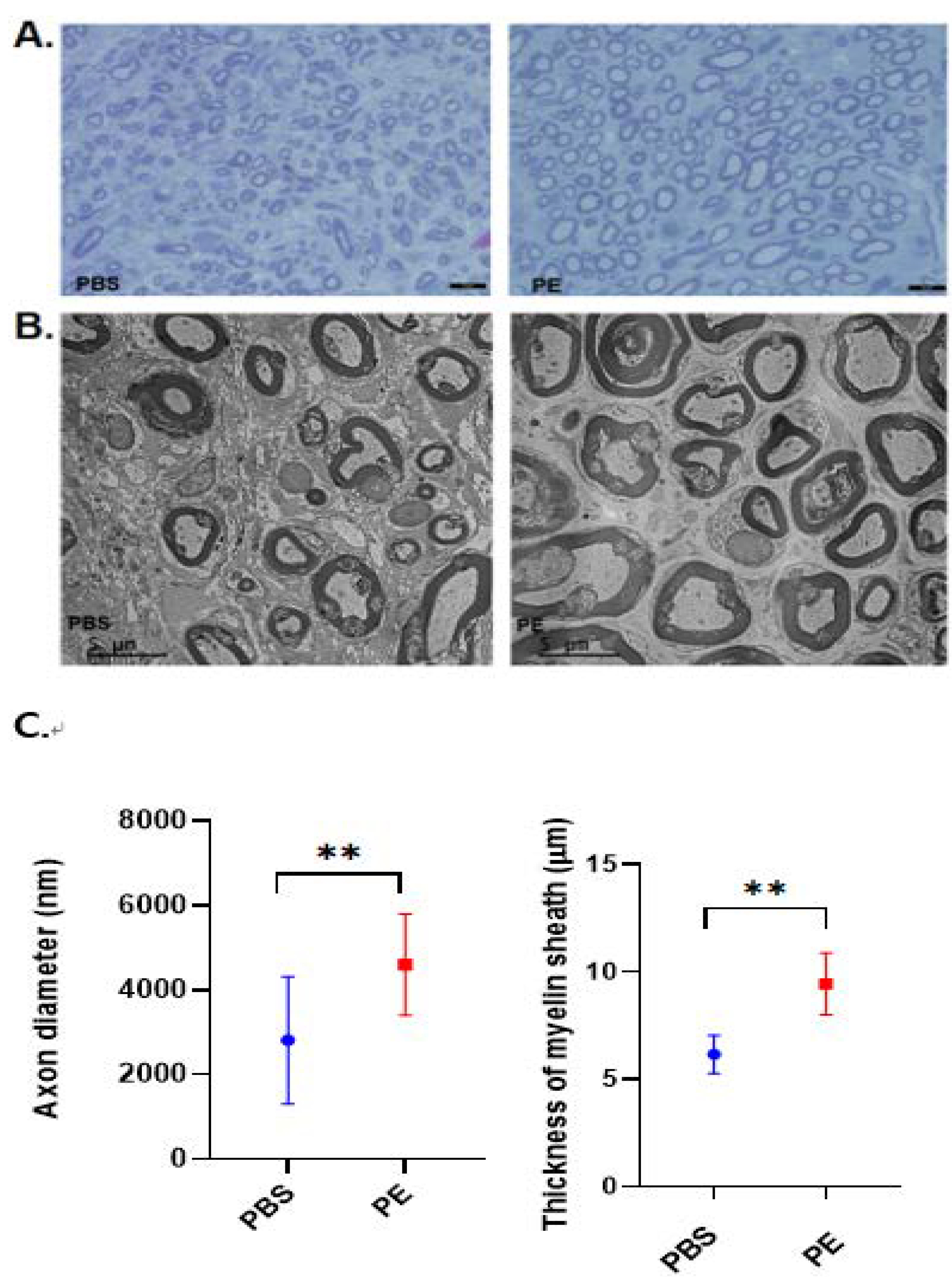
Fig. 10. Crushed facial nerves treated with PE exhibit larger axons compared to the PBS treated control group (toluidine blue staining of semithin sections; solid arrows, A). The size of the axon in the PE-treated group is larger than those in the control group in the semithin section (A) and TEM (B). The bars represent 5 μm. The axon diameter and myelin thickness were measured using image J and are significantly larger in the PE group, detected using Mann-Whitney test. **p=0.0015 (axon diameter), **p=0.0087 (thickness of myelin sheath) (C).
© Exp Neurobiol