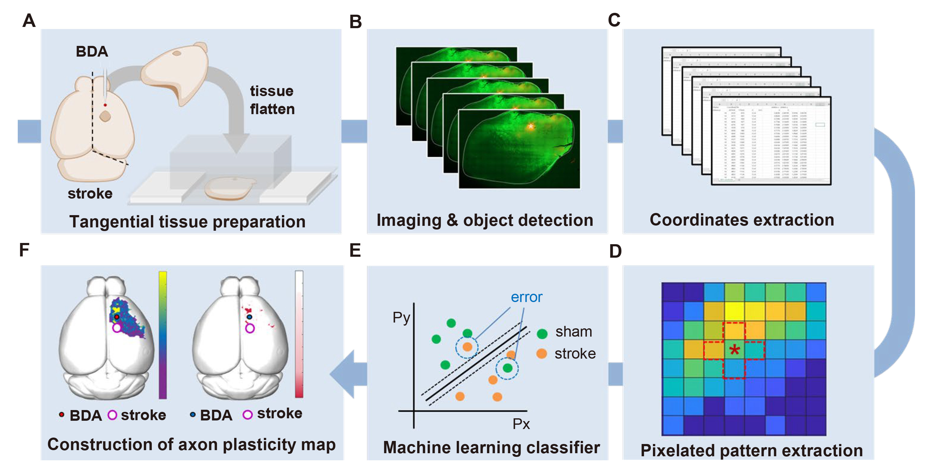
Fig. 1. A schematic workflow of the multi-pixel pattern analysis (MPPA) of the cortical axonal plasticity. (A) Tissue preparation process of BDA injected cortices flattening using customized plexiglass. (B, C) Fluorescence images were subjected to object detection and transposed into Cartesian coordinates. (D) Pixelated coordinates were extracted as a pattern data, (E) and followed machine learning classification to discriminate each pattern into sham or stroke groups. (F) Individual pixel classification accuracy, and accuracy-based statistical significance plotted on 3D brain images.
© Exp Neurobiol