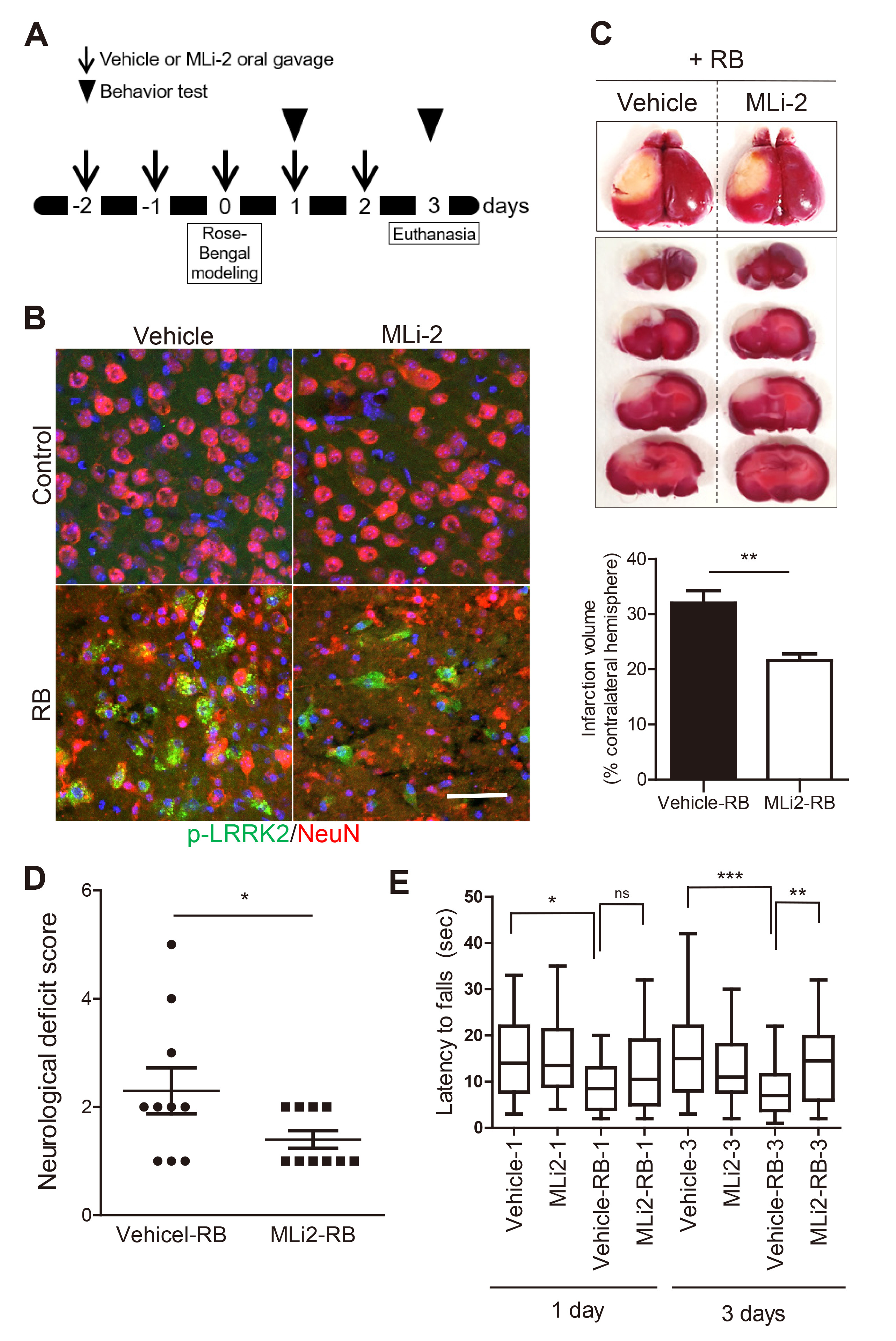
Fig. 2. MLi-2 treatment attenuated the neurological deficits in RB photothrombosis model. (A) Experimental schematic of behavior analysis following the MLi-2 oral gavage. (B) Immunohistochemical analysis of anti-p-LRRK2 (Ser1292; green) and anti-NeuN antibodies (red) at penumbra and the boundary of infarct of brains collected 3 days after RB photothrombosis. Scale bar=25 μm. (C) Representative brain slices stained with TTC and the quantification of TTC-stained cerebral infarct volumes in the vehicle-treated group (Vehicle-RB) and MLi-2-treated group (MLi-2-RB) in RB photothrombosis. Data were expressed as means±SEMs (n=10 per group). **p=0.0033. (D) Neurological deficit scores evaluated 3 days after RB photothrombosis. Data were expressed as means±SEMs (n=10 per group). *p=0.0313, Vehicle-RB group vs. MLi-2-RB group. (E) Wire Hang tests were evaluated 1 day and 3 days after RB photothrombosis. Data were expressed as means±SEMs (n=30, 3 replicates, 10 mice per group). *p<0.5, **p<0.01, ***p<0.001 Vehicle group, MLi-2 treated group, Vehicle-RB group, MLi-2-RB group.
© Exp Neurobiol