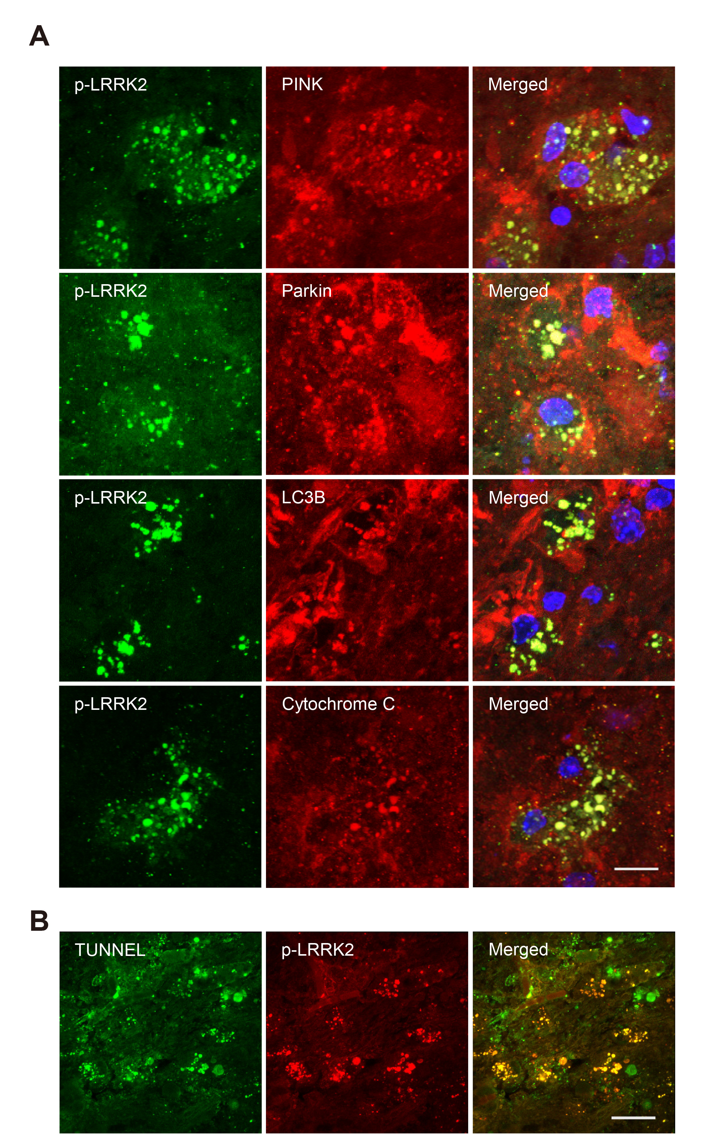
Fig. 3. Colocalization of p-LRRK2 (Ser1292) with of mitophagy-related proteins in the damaged cortex of mice 3 days after RB photothrombosis. (A) Representative confocal microscope images of p-LRRK2 (green) and PINK/Parkin/LC3B/cytochrome c (red) in tissue collected 3 days after RB photothrombosis. Scale bar=25 μm. (B) Representative images of p-LRRK2/TUNEL stained penumbra (area of less damage) of brains collected 3 days after RB photothrombosis. Scale bar=100 μm.
© Exp Neurobiol