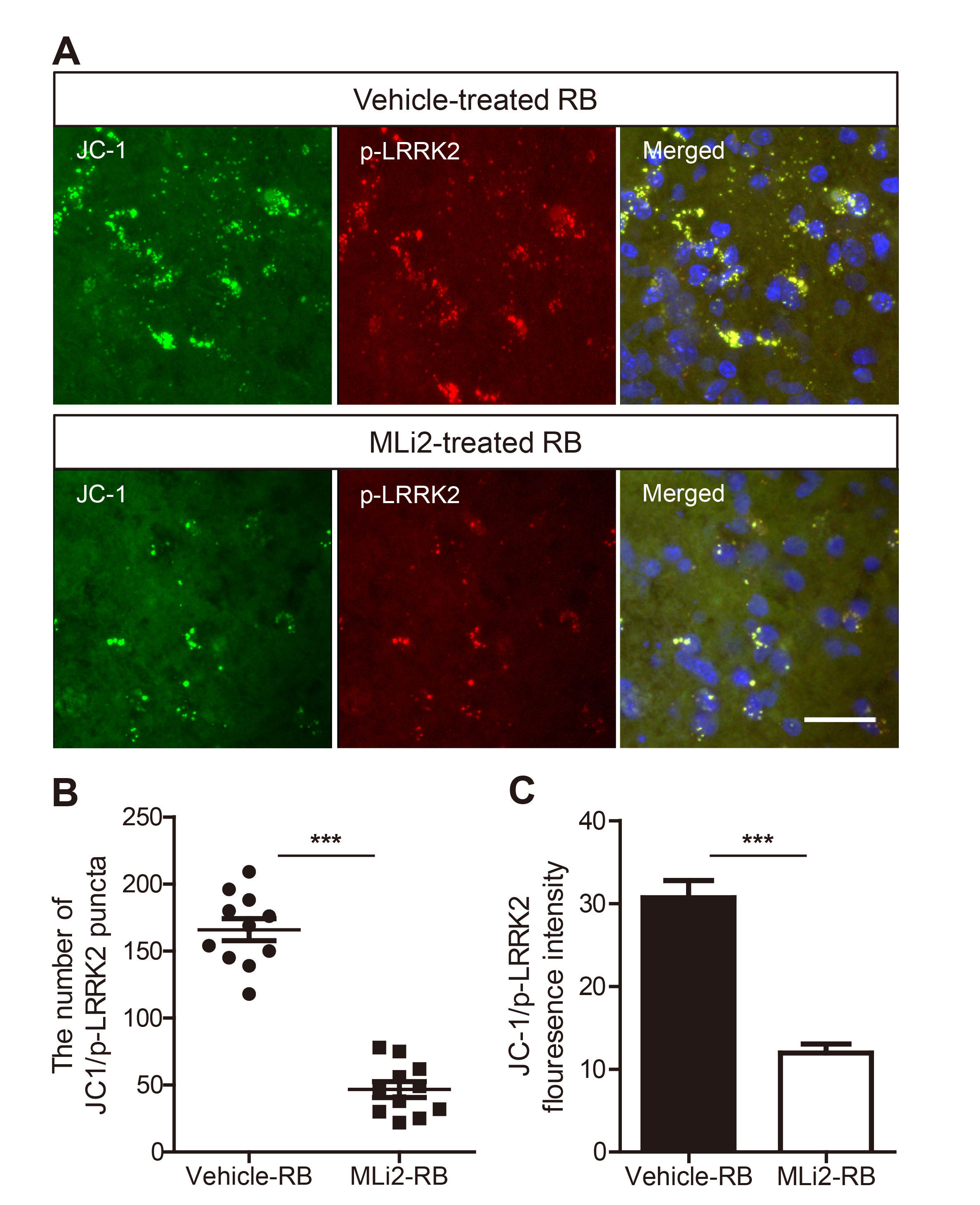
Fig. 4. MLi-2 treatment attenuated mitochondrial apoptosis in RB photothrombosis. (A) Immunohistochemical analysis of anti-p-LRRK2 (S1292; red) and JC-1 dye (mitochondrial marker; green) in tissue collected 3 days after RB photothrombosis. Scale bar=25 μm. (B) The number of JC-1 and p-LRRK2-positive puncta were counted in the confocal microscope images. Data were expressed as means±SEMs (n=10 per group). ***p<0.0001. (C) Graphs represented the mean of the fluorescence intensity of JC-1 and p-LRRK2-positive signal measured by Image J. Data were expressed as means ±SEMs (n=10 per group). ***p<0.0001.
© Exp Neurobiol