Articles

Article Tools
 View Full Text View Full Text |
 Abstract Abstract |
 Article as PDF Article as PDF |
 Print this Article Print this Article |
 Pubmed Pubmed |
 PMC PMC |
 PubReader PubReader |
 Export to Citation Export to Citation |
 Email Alerts Email Alerts |
 Open Access Open Access |
Share this article on :


Stats or Metrics
Article
Original Article
Exp Neurobiol 2011; 20(2): 116-122
Published online June 30, 2011
https://doi.org/10.5607/en.2011.20.2.116
© The Korean Society for Brain and Neural Sciences
Interactions of Dopamine D1 and N-methyl-D-Aspartate Receptors Are Required for Acute Cocaine-Evoked Nitric Oxide Efflux in the Dorsal Striatum
Dong Kun Lee1, Sung Min Ahn1, Yoon-Bo Shim2, Wei Choon Alvin Koh2, Insop Shim3 and Eun Sang Choe1*
Departments of 1Biological Sciences, and 2Chemistry, Pusan National University, Busan 609-735, 3Lab of Neuroscience, AMSRC, Kyung Hee University, Seoul 130-701, Korea
Correspondence to: *To whom correspondence should be addressed.
TEL: 82-51-510-2272, FAX: 82-51-581-2962
e-mail: eschoe@pusan.ac.kr
Alterations in nitric oxide (NO) release in response to psychostimulants in the striatum cause a plastic change contributing to the development and expression of addiction. In this study, regulation of NO efflux evoked by acute cocaine in the dorsal striatum was investigated using real-time detection of NO in vivo. We found that acute systemic injection of cocaine (20 mg/kg) increased NO efflux, which was reduced by the intrastriatal infusion of the dopamine D1 receptor antagonist, SCH23390 (7.5 nmol), and the dopamine D2 receptor agonist, quinpirole (5 nmol). Increased levels of NO efflux by acute cocaine were also reduced by the intrastriatal infusion of the
Keywords: addiction, caudate-putamen, dopamine, glutamate, psychostimulant
Cocaine, an indirect dopamine receptor agonist, is a powerful agent regulating dopaminergic and glutamatergic transmission in the central nervous system. In the striatum, acute exposure to cocaine regulates dopamine release by blocking the reuptake of dopamine in the terminal of the neurons. In addition to dopamine release, repeated exposure to cocaine regulates glutamate release and increases extracellular levels of the transmitter (Rahman et al., 2005). This regulation is mediated through trans-synaptic circuits in the basal ganglia, while it also depends on the responsitivity of several intracellular signaling molecules to cocaine (Kalivas et al., 2003). Increased levels of extracellular dopamine and glutamate as a result of repeated cocaine administration interact with dopamine and glutamate receptors, respectively, that are integrated to the activation of
Neuronal nitric oxide synthase (nNOS) is structurally associated with the cytosolic domain of NMDA receptors in the cerebellum (Christopherson et al., 1999). Stimulation of NMDA receptors activates Ca2+ signaling cascades leading to the activation of calcium/calmodulin-dependent protein kinase (CaMK) and protein phosphatases, which in turn causes nitric oxide (NO) production via alteration of nNOS activity in rat cortical neurons and the dorsal striatum (Nakane et al., 1991; Hayashi et al., 1999). A previous study demonstrated that stimulation of dopamine D1 receptors upregulates NO efflux via nNOS activation in the rat dorsal striatum (Sammut et al., 2006). The increase in NO efflux in the dorsal striatum was also observed after repeated cocaine administration via stimulation of dopamine D1 and group I metabotropic glutamate receptors (mGluRs) (Lee et al., 2010). Stimulation of group I mGluRs was found to potentiate NMDA receptor-mediated NO production (Lee et al., 2010). Collectively, these findings suggest that stimulation of dopamine and/or glutamate receptors after cocaine administration is necessary for the production of NO efflux in the basal forebrain. In addition to repeated cocaine-evoked NO release, it was hypothesized that acute injection of cocaine can control extracellular NO release via dopamine and NMDA receptor stimulation in the dorsal striatum, a key structure participated in extrapyramidal motor and reward pathways. In this study we investigated this hypothesis at receptor levels using real-time detection of NO efflux using a biosensor in the rat dorsal striatum following acute cocaine injection.
MATERIALS AND METHODS
Adult male Sprague Dawley rats (250~300 g) were obtained from Hyo-Chang Science Co. (Taegu, Korea). Rats were individually housed in a controlled environment during all experimental treatments. Food and water were provided ad libitum and rats were maintained on a 12 h light/dark cycle. On the day of the experiments, injections were given in a home cage in a quiet room to minimize stress to the animals. All animal use procedures were approved by the Institutional Animal Care and Use Committee and performed in accordance with the provisions of the National Institute of Health Guide for the Care and Use of Laboratory Animals.
Rats were anesthetized with intraperitoneal (i.p.) injections of chloral hydrate (6 ml/kg) and placed in a Stoelting stereotaxic apparatus. Under aseptic conditions, a 23-gauge stainless steel guide cannula (0.29 mm inner diameter, 10 mm in length) was implanted 1 mm anterior to the bregma, 2.5 mm right of the midline, and 4 mm below the surface of the skull. The guide cannula was sealed with a stainless steel wire of the same length. The rats were allowed at least 5 days to recover from surgery. On the day of the experiments, the inner steel wire was replaced by a 30-gauge stainless steel injection cannula (0.15 mm inner diameter, 12.5 mm in length) that protruded 2.5 mm beyond the guide cannula. Throughout the experiments, rats were randomly divided into four groups (n=4~5 per group): vehicle + saline, vehicle + cocaine, drug + saline, and drug + cocaine. Drugs were infused unilaterally into the central part of the right dorsal striatum 5 min prior to cocaine or saline injection in a volume of 1 µl at a rate of 0.2 µl/min in freely moving rats. The progress of the injection was monitored by observing the movement of a small air bubble along the length of a precalibrated PE-10 tubing inserted between the injection cannula and a 2.5 µl Hamilton microsyringe. After the completion of the injection, the injector was left in place for an additional 5 min to reduce any possible backflow of the solution along the injection tract. Cocaine (Belgopia, Louvain-La-Neuve, Belgium) was dissolved in physiological saline (0.9% NaCl), and each rat received a single systemic injection of cocaine (20 mg/kg, i.p.). Parallel to each drug injection, dimethylsulfoxide (DMSO)/artificial cerebrospinal fluid (aCSF) containing (mM) 123 NaCl, 0.86 CaCl2, 3.0 KCl, 0.89 MgCl2, 0.50 NaH2PO4, and 0.25 NaH2PO4 aerated with 95% O2/5% CO2, pH 7.2~7.4 or NaCl was injected into either the center of the dorsal striatum or the peritoneum 5 min before cocaine or saline injection in each experiment, as a control. All drugs except cocaine were purchased from Tocris Cookson (Ellisville, MO, USA), and the drug solutions were adjusted to pH 7.2~7.4 with 1 N NaOH, if necessary. Drug concentrations were determined based on previous studies (Ahn et al., 2007; Lee et al., 2008; Kim et al., 2009).
The NO microbiosensor was prepared as previously described (Alvin et al., 2008) and was cleaned by cycling the applied potential between +1.4 and -0.2 V for 10 cycles at a scan rate of 500 mV/s in 0.5 M H2SO4 solution, followed by washing with distilled water. Subsequently, the Pt microelectrode was coated with a conductive polymer (CP) through an electropolymerization reaction with a carboxylic acid group TTCA monomer in a 0.1 M TBAP/CH2Cl2 solution. This was accomplished by cycling the potential between 0.0 and 1.5 V two times at a scan rate of 50 mV/s. The electrode was then washed with CH2Cl2 to remove the excess monomer. The CP-coated Pt microelectrode was immersed for 12 h in 0.1 M phosphate buffered saline (PBS; pH 7.0) containing 20 mM 1-ethyl-3 [3-(dimethylamino)-propyl] carbodimide (EDC) to activate the CP carboxylic acid groups. At this point, the EDC treated CP-modified microelectrode was washed with buffer solution and subsequently incubated for 48 h in 5 mM PBS solution containing 6 mg/mL cytochrome c (cyt c) at 4℃. Using this procedure, cyt c was covalently bound through its amine groups to the carboxylic groups on the poly-5,2':5',2-terthiophene-3'-carboxylic acid (poly-TTCA), thus forming amide bonds. The cyt c/poly-TTCA microelectrode was dipped in 1% Nafion solution (diluted with ethanol) for 2 min. The Nafion film was then dried for 1 h in a calcium chloride atmosphere. Nafion films were dried in a low-humidity atmosphere provided by calcium chloride pellicles in a sealed container, which increased stability.
The real-time detection of NO was performed previously described (Alvin et al., 2008; Lee et al., 2010). Briefly, rats were anesthetized with 8% chloral hydrate and placed in a Stoelting stereotaxic apparatus. The enzyme-coated electrode and the reference electrode of the NO microbiosensor were inserted under ascetic conditions at the coordinates of 1 mm anterior of the bregma, 2.5 mm right of the midline, and 4 mm below the surface of the skull where the drugs were infused. The sensor electrode was connected to a Potentiostat/Galvanostat (Model PT-1; Kosentech, Pusan, Korea), which measured and recorded the electrochemical signal. The sensor was calibrated with a series of NO standard solutions during pre- and post experimental measurements. A 20-min time point was selected based on a time course of cocaine injection with peak NO levels at 20 min.
Data from NO measurements were recorded as currents which were converted to concentrations (µM). Statistical significance between groups was determined using one-way ANOVA followed by a Tukey's honestly significant difference test in GraphPad Prism 4 (GraphPad Software, San Diego, CA, USA). The level of statistical significance was set to p < 0.05.
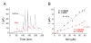
The NO microbiosensor was calibrated before and after measurement of NO levels in the series of experiments (Fig. 1A). Under optimized conditions, the steady-state currents exhibited a linear relationship with NO concentrations in the range of 0~55.0 µM (Fig. 1B). The sensitivity of the NO microbiosensor was 0.117 ± 0.006 µA/µM. The detection limit of NO concentration was 13 ± 3 nM based on five replicates to determine the standard deviation of the blank noise (95% confidence level,
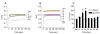
To determine whether acute injection of cocaine alters the NO efflux in the dorsal striatum, we measured real-time amperometric NO responses for 120 s before and 5 min after acute saline or cocaine injection up to 60 min at 10 min time intervals. Acute systemic injection of cocaine increased NO levels compared with saline injection (Fig. 2A and 2B). Semiquantitation confirmed that as a result of cocaine, but not saline, injection, NO levels were significantly increased at 10 min, remained up to 20 min, and then returned to basal levels (Fig. 2C).
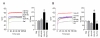
Because acute cocaine increased NO levels, this experiment was conducted to determine the involvement of dopamine receptors in the regulation of NO efflux in the dorsal striatum. Intrastriatal infusion of the dopamine D1 receptor antagonist, SCH23390 (7.5 nmol), significantly decreased an acute cocaine-evoked increase in NO levels (Fig. 3A). Intrastriatal infusion of the dopamine D2 receptor agonist, quinpirole (5 nmol), also significantly decreased NO levels (Fig. 3B). However, SCH23390 or quinpirole alone did not alter NO levels compared with the saline-injected group (Fig. 3).
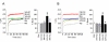
Because dopamine D1 receptor stimulation interacts with NMDA receptors in the ventral tegmatal area (Schilström et al., 2006), this experiment was conducted to determine the involvement of NMDA receptors in the regulation of NO efflux after acute cocaine administration. Intrastriatal infusion of the NMDA receptor antagonists, MK801 (2 nmol) and AP5 (2 nmol), significantly reduced the acute cocaine-evoked increase in NO levels (Fig. 4). However, MK801 or AP5 alone did not alter NO levels compared with the saline-injected group (Fig. 4). Changes in real-time NO levels caused by the drug injection in all experiments are summarized in Table 1.
The present data demonstrate that acute injection of cocaine increases NO efflux in the dorsal striatum which is likely regulated by the interactions of dopamine D1 and NMDA receptors. Although evidence concerning the mechanisms underlying NO efflux after exposure to psychostimulants is limited, the present finding implies that integration of dopamine signals to NMDA receptors in the dorsal striatum is required for NO efflux.
A previous study demonstrated that striatal NO efflux induced by electrical or pharmacological stimuli of the substantia nigra is regulated by dopamine D1 receptor stimulation (Sammut et al., 2006), suggesting that NO release in the striatum is primarily mediated by the stimulation of dopamine D1 receptors. Similarly, the present data demonstrated that increased levels of NO efflux in the dorsal striatum after acute injection of cocaine are decreased by blockade of dopamine D1 receptors or stimulation of dopamine D2 receptors. It is well known that dopamine D1 receptor is coupled to excitatory G-protein (Gs) and stimulation of the receptor increases the levels of adenylyl cyclase (AC), which in turn activates (phosphorylates) NMDA receptors via protein kinase A (PKA)-dependent or independent cAMP activity (Cepeda et al., 1998; Cepeda and Levine, 1998; Scott et al., 2002). Thus, it can be postulated that NMDA receptors can be activated by the stimulation of dopamine D1 receptors after acute cocaine administration, which plays a critical role in NO efflux into the dorsal striatum. This speculation is supported by the finding that blockade of either dopamine D1 or NMDA receptors abolished the increase in NO efflux in the dorsal striatum after repeated exposure to cocaine (Lee et al., 2010). Similar to this finding, systemic injection of the NMDA receptor antagonist, MK801, decreased striatal NO efflux evoked by electrical stimuli of the frontal cortex (Sammut et al., 2007). Taken together, these findings indicate that NMDA receptor stimulation may couple dopamine signals to Ca2+ cascades for the regulation of NO efflux after acute cocaine administration.
Alterations in Ca2+ levels by the stimulation of NMDA receptors consequently cause nNOS activation followed by NO production via multiple steps of protein kinase and phosphatase interactions (Lee et al., 2010). Previous studies demonstrated that stimulation of dopamine D1 receptors activates AC/cAMP (or PKA) cascades and phosphorylates nNOS (Centonze et al., 2001; Yu et al., 2002; Park and West, 2009). Activation of protein phosphatases by NMDA receptor-stimulated Ca2+-cascades in the striatum upregulates NO efflux via dephosphorylation of phosphorylated nNOS (Nishi et al., 2005). Collectively, these findings suggest that dopamine D1 receptor-dependent Ca2+ influx via NMDA receptor stimulation after acute injection of cocaine is required for the regulation of NO efflux in the dorsal striatum. It is important to note that repeated cocaine synergistically activates NMDA receptors via stimulation of dopamine and group I mGluRs, while in acute cocaine only dopamine signals contribute to activate NMDA receptors for the production of NO efflux in the dorsal striatum (Choe et al., 2011).
NO has been suggested as a retrograde neurotransmitter that may diffuse from the postsynaptic neurons to the presynaptic neurons (Snyder, 1992). Thus, dopamine receptors in GABAergic neurons stimulated by acute cocaine may increase the production of NO and then increase to diffuse it into the presynaptic dopamine terminals of the dorsal striatum (Lee et al., unpublished observations). For instance, NOS inhibitors blocked cocaine or methamphetamine-induced dopamine releases in the striatum (Bowyer et al., 1995; Inoue et al., 1996), suggesting that inhibition of NO formation reduces the development of locomotor activity by the modulation of dopamine releases or activation of postsynaptic dopamine receptors in the striatum. Taken together, these results strongly suggest that activation of dopamine receptors has an important role in NO-mediated behavioral effects probably via NMDA receptor stimulation produced by acute cocaine administration. On the other hand, this difference in the regulation of NO efflux after cocaine administration (acute vs. repeated) may results in the expression of behavioral sensitization.
In summary, regulation of acute cocaine-evoked NO efflux was determined by using real-time detection of NO efflux in the dorsal striatum. We found that acute systemic injection of cocaine upregulates NO levels via dopamine D1 receptor-stimulated NMDA receptors. Like repeated exposure to cocaine, NO efflux can be upregulated by acute cocaine and interactions of dopamine D1 receptors and NMDA receptors can contribute to this upregulation in the dorsal striatum.
Table 1. Changes in real-time NO values caused by the drug injection following acute exposure to cocaine in the dorsal striatum
Values represent mean ± SEM (µM). * and # represent an increase and a decrease in the NO levels compared with acute saline and cocaine administration, respectively.
- Ahn SM, Kim SW, Choe ES. Cocaine increases immunoglobulin heavy chain binding protein and caspase-12 expression in the rat dorsal striatum. Psychopharmacology (Berl) 2007;195:407-414.

- Alvin Koh WC, Rahman MA, Choe ES, Lee DK, Shim YB. A cytochrome c modified-conducting polymer microelectrode for monitoring in vivo changes in nitric oxide. Biosens Bioelectron 2008;23:1374-1381.
- Bowyer JF, Clausing P, Gough B, Slikker W, Holson RR. Nitric oxide regulation of methamphetamine-induced dopamine release in caudate/putamen. Brain Res 1995;699:62-70.
- Centonze D, Picconi B, Gubellini P, Bernardi G, Calabresi P. Dopaminergic control of synaptic plasticity in the dorsal striatum. Eur J Neurosci 2001;13:1071-1077.
- Cepeda C, Colwell CS, Itri JN, Chandler SH, Levine MS. Dopaminergic modulation of NMDA-induced whole cell currents in neostriatal neurons in slices: contribution of calcium conductances. J Neurophysiol 1998;79:82-94.
- Cepeda C, Levine MS. Dopamine and N-methyl-D-aspartate receptor interactions in the neostriatum. Dev Neurosci 1998;20:1-18.
- Choe ES, Ahn SM, Yang JH, Go BS, Wang JQ. Linking cocaine to endoplasmic reticulum in striatal neurons: role of glutamate receptors. Basal Ganglia 2011
- Christopherson KS, Hillier BJ, Lim WA, Bredt DS. PSD-95 assembles a ternary complex with the N-methyl-D-aspartic acid receptor and a bivalent neuronal NO synthase PDZ domain. J Biol Chem 1999;274:27467-27473.
- Go BS, Ahn SM, Shim I, Choe ES. Activation of c-Jun N-terminal kinase is required for the regulation of endoplasmic reticulum stress response in the rat dorsal striatum following repeated cocaine administration. Neuropharmacology 2010;59:100-106.
- Hayashi Y, Nishio M, Naito Y, Yokokura H, Nimura Y, Hidaka H, Watanabe Y. Regulation of neuronal nitric-oxide synthase by calmodulin kinases. J Biol Chem 1999;274:20597-20602.
- Inoue H, Arai I, Shibata S, Watanabe S. NG-nitro-L-arginine methyl ester attenuates the maintenance and expression of methamphetamine-induced behavioral sensitization and enhancement of striatal dopamine release. J Pharmacol Exp Ther 1996;277:1424-1430.
- Kalivas PW, McFarland K, Bowers S, Szumlinski K, Xi ZX, Baker D. Glutamate transmission and addiction to cocaine. Ann N Y Acad Sci 2003;1003:169-175.
- Kim SM, Ahn SM, Go BS, Wang JQ, Choe ES. Alterations in AMPA receptor phosphorylation in the rat striatum following acute and repeated cocaine administration. Neuroscience 2009;163:618-626.
- Lee DK, Bian S, Rahman MA, Shim YB, Choe ES. Repeated cocaine administration increases N-methyl-d-aspartate NR1 subunit, extracellular signal-regulated kinase and cyclic AMP response element-binding protein phosphorylation and glutamate release in the rat dorsal striatum. Eur J Pharmacol 2008;590:157-162.
- Lee DK, Koh WC, Shim YB, Shim I, Choe ES. Repeated cocaine administration increases nitric oxide efflux in the rat dorsal striatum. Psychopharmacology (Berl) 2010;208:245-256.
- Nakane M, Mitchell J, Forstermann U, Murad F. Phosphorylation by calcium calmodulin-dependent protein kinase II and protein kinase C modulates the activity of nitric oxide synthase. Biochem Biophys Res Commun 1991;180:1396-1402.
- Nishi A, Watanabe Y, Higashi H, Nairn AC, Greengard P. Glutamate regulation of DARPP-32 phosphorylation in neostriatal neurons involves activation of multiple signaling cascades. Proc Natl Acad Sci USA 2005;102:1199-1204.
- Park DJ, West AR. Regulation of striatal nitric oxide synthesis by local dopamine and glutamate interactions. J Neurochem 2009;111:1457-1465.
- Rahman MA, Kwon NH, Won MS, Choe ES, Shim YB. Functionalized conducting polymer as an enzyme immobilizing substrate: an amperometric glutamate microbiosensor for in vivo measurements. Anal Chem 2005;77:4854-4860.
- Sammut S, Dec A, Mitchell D, Linardakis J, Ortiguela M, West AR. Phasic dopaminergic transmission increases NO efflux in the rat dorsal striatum via a neuronal NOS and a dopamine D(1/5) receptor-dependent mechanism. Neuropsychopharmacology 2006;31:493-505.
- Sammut S, Park DJ, West AR. Frontal cortical afferents facilitate striate nitric oxide transmission in vivo via a NMDA receptor and neuronal NOS-dependent mechanism. J Neurochem 2007;103:1145-1156.
- Schilström B, Yaka R, Argilli E, Suvarna N, Schumann J, Chen BT, Carman M, Singh V, Mailliard WS, Ron D, Bonci A. Cocaine enhances NMDA receptor-mediated currents in ventral tegmental area cells via dopamine D5 receptor-dependent redistribution of NMDA receptors. J Neurosci 2006;26:8549-8558.
- Scott L, Kruse MS, Forssberg H, Brismar H, Greengard P, Aperia A. Selective up-regulation of dopamine D1 receptors in dendritic spines by NMDA receptor activation. Proc Natl Acad Sci USA 2002;99:1661-1664.
- Snyder SH. Nitric oxide: first in a new class of neurotransmitters. Science 1992;257:494-496.
- Yu J, Yu L, Chen Z, Zheng L, Chen X, Wang X, Ren D, Zhao S. Protein inhibitor of neuronal nitric oxide synthase interacts with protein kinase A inhibitors. Mol Brain Res 2002;99:145-149.