Articles

Article Tools
 View Full Text View Full Text |
 Abstract Abstract |
 Article as PDF Article as PDF |
 Print this Article Print this Article |
 Pubmed Pubmed |
 PMC PMC |
 PubReader PubReader |
 Export to Citation Export to Citation |
 Email Alerts Email Alerts |
 Open Access Open Access |
Share this article on :


Stats or Metrics
Article
Original Article
Exp Neurobiol 2018; 27(6): 593-604
Published online December 5, 2018
https://doi.org/10.5607/en.2018.27.6.593
© The Korean Society for Brain and Neural Sciences
Characterization of Tetrodes Coated with Au Nanoparticles (AuNPs) and PEDOT and Their Application to Thalamic Neural Signal Detection in vivo
Daae Lee1, Hyeong Cheol Moon2,3, Bao-Tram Tran3, Dae-Hyuk Kwon4, Yong Hee Kim5, Sang-Don Jung5, Jong Hoon Joo1, and Young Seok Park2,3*
1Department of Advanced Materials Engineering, Chungbuk National University, Cheongju 28644, Korea.
2Department of Neurosurgery, Chungbuk National University Hospital, Cheongju 28644, Korea.
3Department of Neurosurgery, Chungbuk National University, Cheongju 28644, Korea.
4Neuroscience Research Institute, Brain-Bio center, University of Suwon, Hwaseong 18323, Korea.
5Synaptic Devices Research Section, Electronics and Telecommunications Research Institute, Daejeon 34129, Korea.
Correspondence to: *To whom correspondence should be addressed.
TEL: 82-43-269-6080, FAX: 82-43-273-1614
e-mail: youngseokparkmd@gmail.com
Abstract
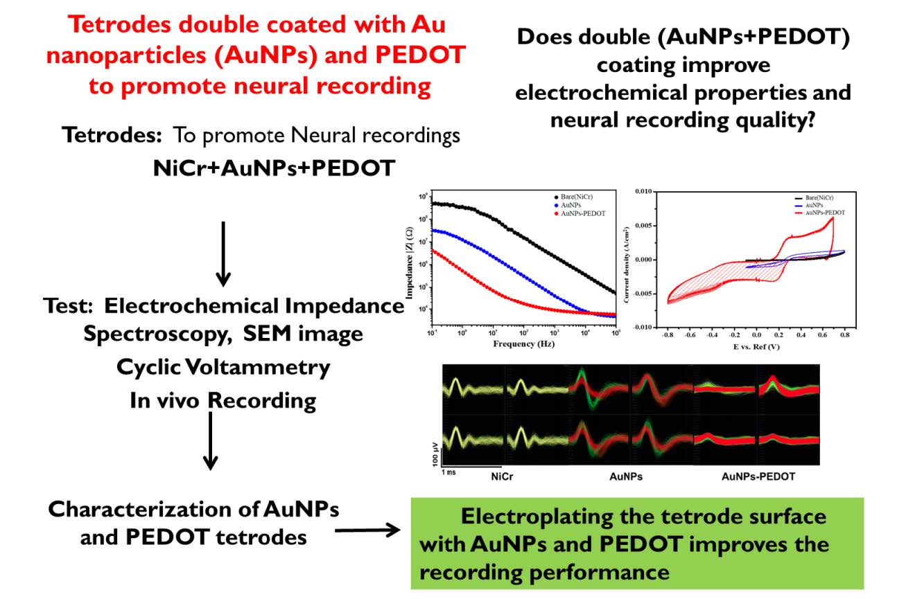
Tetrodes, consisting of four twisted micro-wires can simultaneously record the number of neurons in the brain. To improve the quality of neuronal activity detection, the tetrode tips should be modified to increase the surface area and lower the impedance properties. In this study, tetrode tips were modified by the electrodeposition of Au nanoparticles (AuNPs) and dextran (Dex) doped poly (3,4-ethylenedioxythiophene) (PEDOT). The electrochemical properties were measured using electrochemical impedance spectroscopy (EIS) and cyclic voltammetry (CV). A decrease in the impedance value from 4.3 MΩ to 13 kΩ at 1 kHz was achieved by the modified tetrodes. The cathodic charge storage capacity (CSCC) of AuNPs-PEDOT deposited tetrodes was 4.5 mC/cm2, as determined by CV measurements. The tetrodes that were electroplated with AuNPs and PEDOT exhibited an increased surface area, which reduced the tetrode impedance.
Graphical Abstract
Keywords: microelectrodes, electrodes, Implanted, neuron, Signal detection, Thalamus