Articles

Article Tools
 View Full Text View Full Text |
 Abstract Abstract |
 Article as PDF Article as PDF |
 Print this Article Print this Article |
 Pubmed Pubmed |
 PMC PMC |
 PubReader PubReader |
 Export to Citation Export to Citation |
 Email Alerts Email Alerts |
 Open Access Open Access |
Share this article on :


Stats or Metrics
Article
Original Article
Exp Neurobiol 2020; 29(1): 50-69
Published online February 7, 2020
https://doi.org/10.5607/en.2020.29.1.50
© The Korean Society for Brain and Neural Sciences
Spatiotemporal Profile and Morphological Changes of NG2 Glia in the CA1 Region of the Rat Hippocampus after Transient Forebrain Ischemia
Xuyan Jin1,2, Tae-Ryong Riew1, Soojin Kim1, Hong Lim Kim3 and Mun-Yong Lee1,2*
1Department of Anatomy, Catholic Neuroscience Institute, College of Medicine, The Catholic University of Korea, 2Department of Biomedicine and Health Sciences, College of Medicine, The Catholic University of Korea, 3Integrative Research Support Center, Laboratory of Electron Microscope, College of Medicine, The Catholic University of Korea, Seoul 06591, Korea
Correspondence to: *To whom correspondence should be addressed.
TEL: 82-2-2258-7261, FAX: 82-2-536-3110
e-mail: munylee@catholic.ac.kr
This is an Open Access article distributed under the terms of the Creative Commons Attribution Non-Commercial License
(http://creativecommons.org/licenses/by-nc/4.0) which permits unrestricted non-commercial use, distribution, and
reproduction in any medium, provided the original work is properly cited.
Abstract
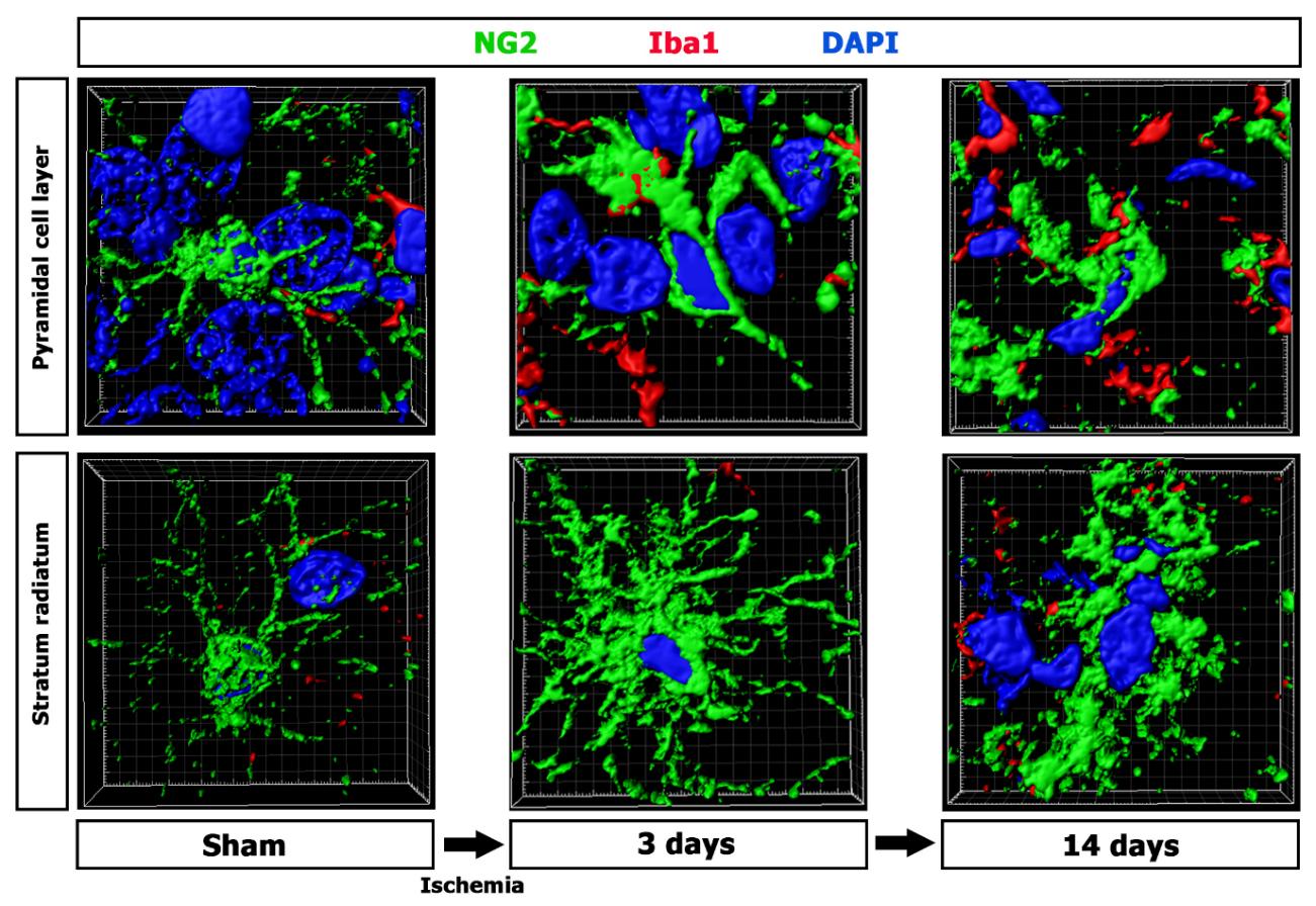
Neuron-glial antigen-2 (NG2) glia undergo proliferation and morphological changes following brain insults. Here, we show that NG2 glia is activated in a characteristic time- and layer-specific manner in the ischemia-vulnerable CA1 region of the rat hippocampus. Resting NG2 glia of the pyramidal cell layer (somatic region) shared morphological features with those of the neighboring dendritic stratum radiatum. During the postischemic period, reactive NG2 glia of the pyramidal cell layer exhibited shortened, scarcely branched processes, while those of the stratum radiatum had multiple branching processes with their arborization being almost indiscernible 7~14 days after reperfusion. Immunoelectron microscopy demonstrated that NG2 immunoreactivity was specifically associated with the plasma membrane and the adjacent extracellular matrix of NG2 glia in the stratum radiatum at 14 days. NG2 glia also exhibited differences in their numbers and proliferation profiles in the two examined hippocampal strata after ischemia. In addition, induced NG2 expression in activated microglia/macrophages exhibited a characteristic strata-dependent pattern in the ischemic CA1 hippocampus. NG2 induction was prominent in macrophage-like phenotypes which were predominantly localized in the pyramidal cell layer, compared with activated stellate microglial cells in the stratum radiatum. Thus, our data demonstrate that activation of NG2 glia and the induction of NG2 expression in activated microglia/macrophages occur in a distinct time- and layer-specific manner in the ischemic CA1 hippocampus. These characteristic profiles of reactive NG2 glia could be secondary to the degeneration processes occurring in the cell bodies or dendritic domains of hippocampal CA1 pyramidal neurons after ischemic insults.
Graphical Abstract
Keywords: Hippocampus, Microglia, NG2 glia, Pyramidal cell layer, Stratum radiatum