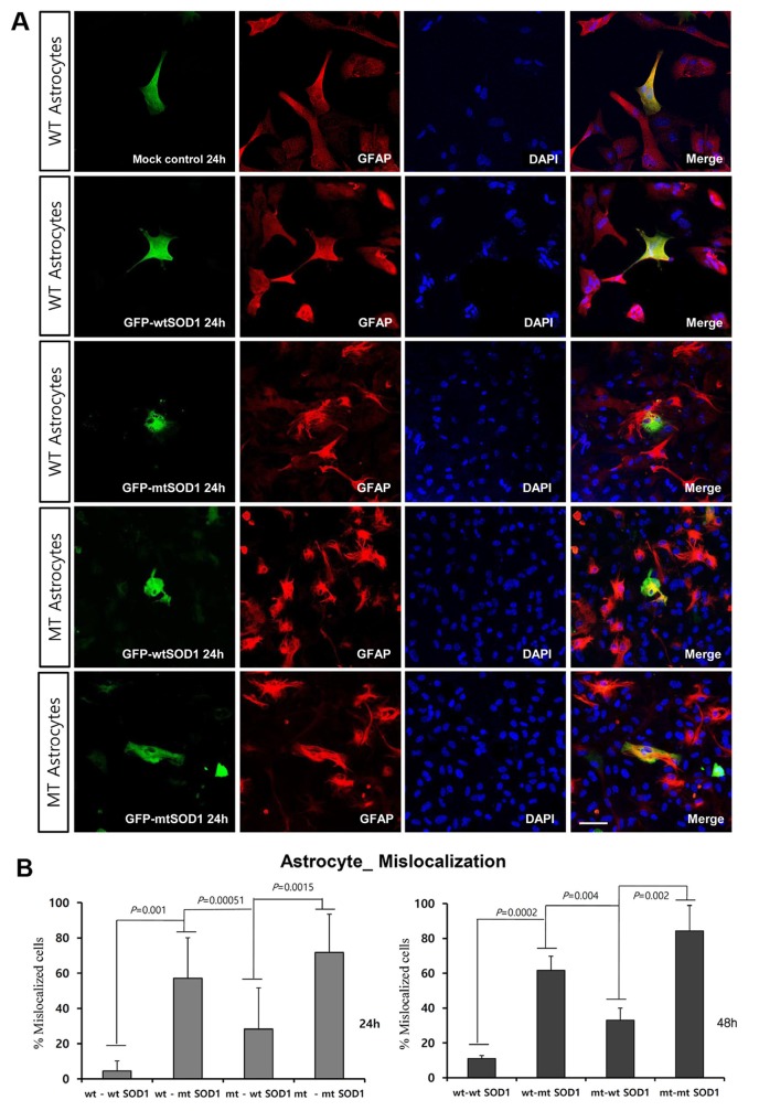
Fig. 2. Intracellular localization of transfected GFP-wt or mtSOD1 in cultured primary astrocytes from G93A-SOD1 mice. (A) Astrocytes stained with GFAP (red) and DAPI (blue) nuclear counterstain. Mock transfection did not affect the cellular localization in both WT and MT astrocytes. In WT astrocytes transfected with GFP-wtSOD1, protein predominantly colocalized in the cytoplasm and the nucleus, whereas in those transfected with mtSOD1, protein localized in the cytosol. Scale bar=50 µm. (B) Cytoplasmic mislocalization was examined by measuring the intensity of hSOD1-EGFP fluorescence at 24 h and 48 h post-transfection. Data are expressed as mean values (±standard error).
© Exp Neurobiol