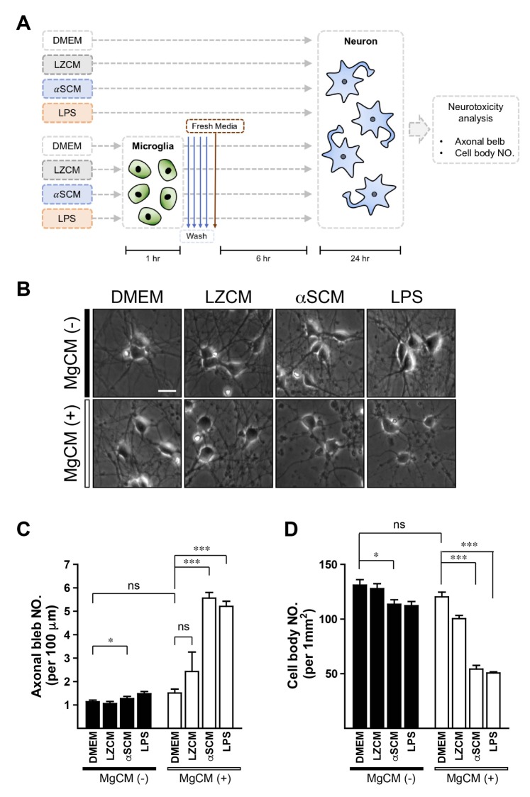
Fig. 1. Microglial neurotoxicity by neuronreleased α-synuclein. Rat primary neurons were treated with various types of conditioned medium for 24 hours. (A) Experimental scheme. (B) Representative images; DMEM, LZCM, αSCM, and LPS treated neurons (upper panels) and DMEM-MgCM, LZCM-MgCM, αSCM-MgCM, and LPS-MgCM treated neurons (lower panels). (C) The numbers of axonal blebs formations. (D) The loss of neuronal cell bodies. All data were analyzed by one-way ANOVA. Error bars represent the s.e.m. ns; not significant; *p<0.05; ***p<0.001. Scale bar, 10 µm.
© Exp Neurobiol