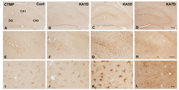
Fig. 1. Representative photograph with immunohistochemical staining with anti-CTMP primary antibodies in kainic acid (KA) treated mice. Mice were administrated with KA for 1 (B, F, J), 3 (C, G, K) and 7 days (D, H, L). Brain sections were collected and stained for CTMP antibody. In the control (A, E, I), CTMP was weakly found in the hippocampus. In the KA-injured hippocampus, a strong staining was observed at 1 day post-lesion and became maximal at 3 days and return to normal level at 7 days. Higher magnification of hippocampus showed sequential changes of CTMP-positive cells. Note that CTMP-positive cells exhibited the star-shaped morphology (I~L). (Scale bars=200 µm in A~D, 100 µm in E~H, 20 µm in I~L).
© Exp Neurobiol