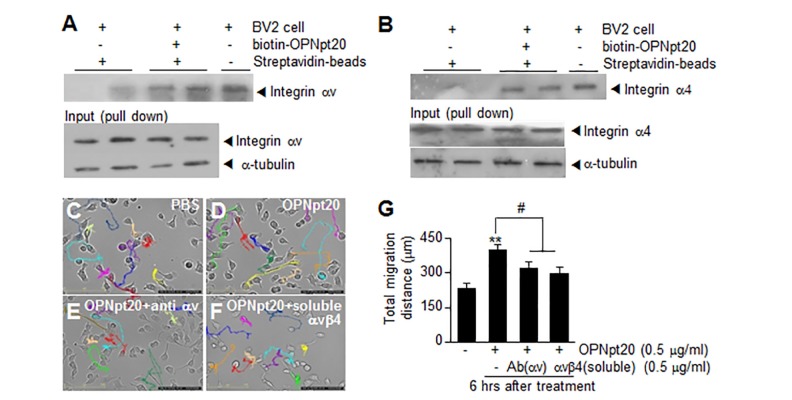
Fig. 3. OPNpt20 binding to αv integrin in BV2 cells. (A, B) Biotinylated-OPNpt20 (1 µg/ml) was incubated with whole cell lysate of BV2 for 6 hrs and pull-down assays were performed using streptavidin agarose beads. Levels of αv integrin (A) or α4 integrin (B) were assessed by immunoblotting. (C~G) BV2 cells were incubated with OPNpt20 for 6 hrs in the presence of anti-αv integrin antibody (0.5 µg/ml) or soluble αvβ3 integrin (0.5 µg/ml). Cell motilities were measured every 10 minutes for 6 hrs by live cell imaging (C~F) and total migration distances were measured using Image J software (G). Results are presented as means±SEMs. **p<0.01 versus PBS-treated controls and #p<0.05 versus OPNpt20-treated cells. Scale bars represent 125 µm (C~F).
© Exp Neurobiol