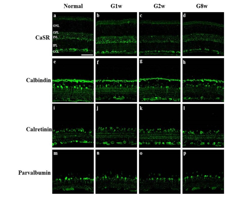
Fig. 5. Confocal microscopic analysis of normal and glaucoma retinas processed for CaSR and calcium-binding proteins immunohistochemistry. Immunohistochemistry for CaSR (a~d), calbindin (e~h), calretinin (i~l) and parvalbumin (m~p) was performed in normal retina (Normal: a, e, i, m), and in 1 week (G1w: b, f, j, n), 2 week (G2w: c, g, k, o), and 8 week (G8w: d, h, l, p) glaucoma retinas. CaSR immunoreactivity in the neurons in the ganglion cell layer (GCL) clearly reduced in G2w and G8w retinas. Calbindin immunoreactivity in amacrine cells in the inner nuclear layer (INL) gradually reduced in glaucoma retinas and that in the axons of ganglion cells increased. Calretinin immunoreactivity in amacrine cells showed no large change in glaucoma retinas. Parvalbumin immunoreactivity in the entire AII amacrine cell profile gradually reduced in glaucoma retinas. IPL, inner plexiform layer; ONL, outer nuclear layer; OPL, outer plexiform layer. Scale bar indicates 50 µm.
© Exp Neurobiol