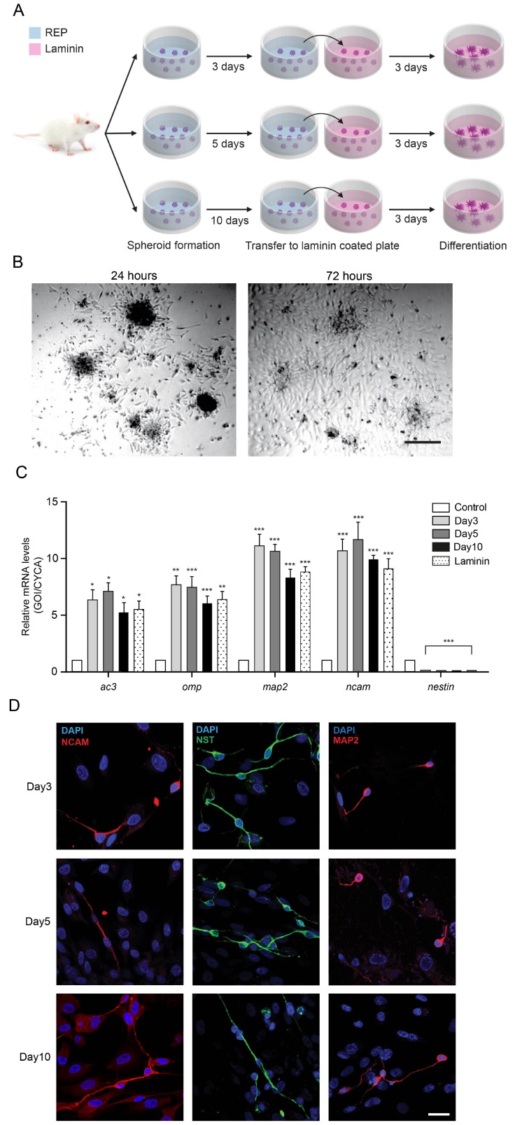
Fig. 2. Differentiation of ORN spheroids and its verification. (A) Illustration of differentiation of ORN spheroids depending on the cultivation period. (B) Microscopic images of plated ORN spheroids on the laminin-pretreated plate for 72 hours (scale bar, 100 µm). (C) qPCR analyses of ORN developmental markers in the ORN spheroids depending on the cultivation time. Control group is plated ORNs on the laminin-coated plate for 72 hours. (D) Immunocytochemistry with anti-NST, anti-NCAM, and anti-MAP2 to identify differentiation of ORNs cultivated for 72 hours (scale bar, 50 µm). Data were obtained from three separate animal culture experiments.
© Exp Neurobiol