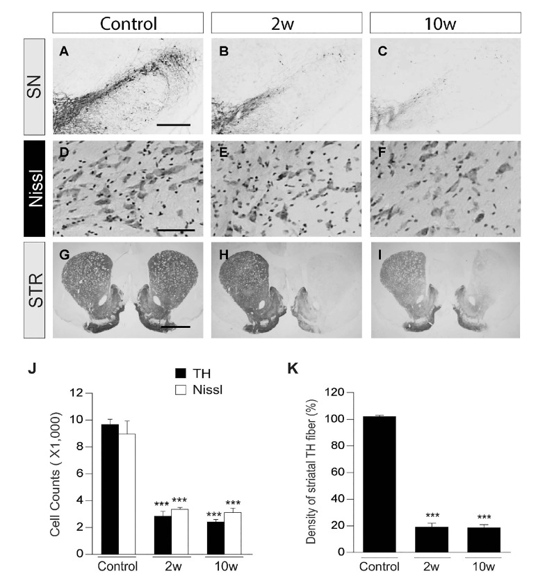
Fig. 1. Degeneration of dopamine neurons is completed at 2 weeks post MPP+in vivo. (A~I) Photomicrographs of TH+ (A~C) and Nissl+ (D~F) cells in the substantia nigra pars compacta (SNpc) (A~F), TH+ fibers in the striatum (STR) (G~I) at 2 (B, E, H) and 10 (C, F, I) weeks (w) after a unilateral medial forebrain bundle (MFB) injection of MPP+ or at 2 w after injection of PBS as a control (A, D, G). Scale bars: 400 µm (A~C, SNpc), 100 µm (D~F, SNpc), 2 mm (G~I, STR). Magnification: 4× (A~C), 10× (D~F), 1.25× (G~I). (J) The number of TH+ and Nissl+ cells in the SNpc. (K) Optical density of TH+ fibers in the STR. J and K, ***p<0.001, significantly different from control. Mean ± s.e.m.; One way ANOVA with Newman-Keuls analysis and Student t-Test.
© Exp Neurobiol