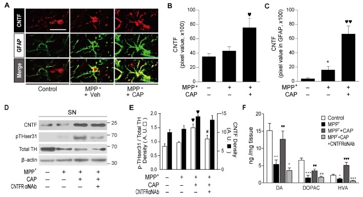
Fig. 4. Delayed CAP treatment increases astrocytic CNTF expression in MPP+-lesioned rat SNpc in vivo. (A) Fluorescence images of CNTF (red) and GFAP (green) and both images are merged in the SNpc of control and in presence of CAP or Veh in the SNpc at 10 weeks (w) post MPP+. Scale bar: 20 µm. Magnification: 20×. Quantification of CNTF (B) or CNTF expression in GFAP+ astrocytes (C). (D and E) Western blot analysis (D), quantification (E) in the rat SN at 10 weeks post MPP+. (F) HPLC analysis showing levels of DA and its metabolites (DOPAC and HVA) in the ipsilateral STR at 10 weeks post MPP+. B, C, E and F, *p<0.05, **p<0.01, ***p<0.001, significantly different from control; ♥p<0.05, ♥♥p<0.01, ♥♥♥p<0.001, significantly different from MPP++Veh; #p<0.05, ##p<0.01, ###p<0.001, significantly different from MPP++CAP. Mean±s.e.m.; One way ANOVA with Newman-Keuls analysis.
© Exp Neurobiol