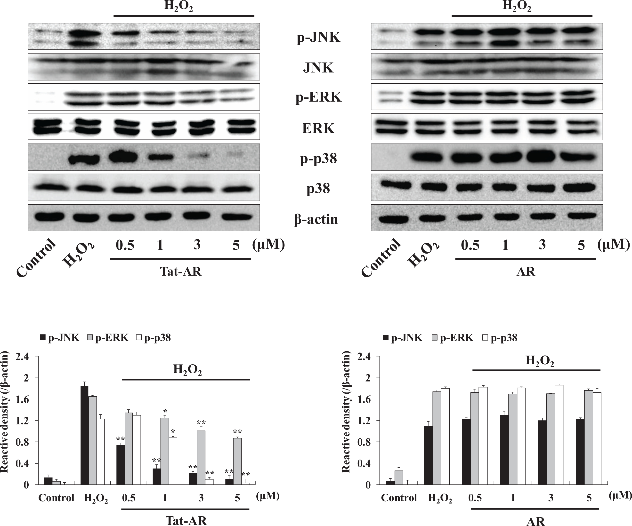
Fig. 4. Effects of Tat-AR protein on H2O2-induced MAPK activation in HT-22 cells. The cells were stimulated with H2O2 (1 mM) for 30 min with or without being pretreated with Tat-AR protein (0.5~5 μM) for 1 h. Then, the cells were prepared and analyzed for phosphorylation of JNK, ERK, p38 levels by Western blotting and the band intensities were measured by densitometer. *p<0.05 and **p<0.01 compared with H2O2-treated cells. Data are repressed as mean±SEM (n=3).
© Exp Neurobiol