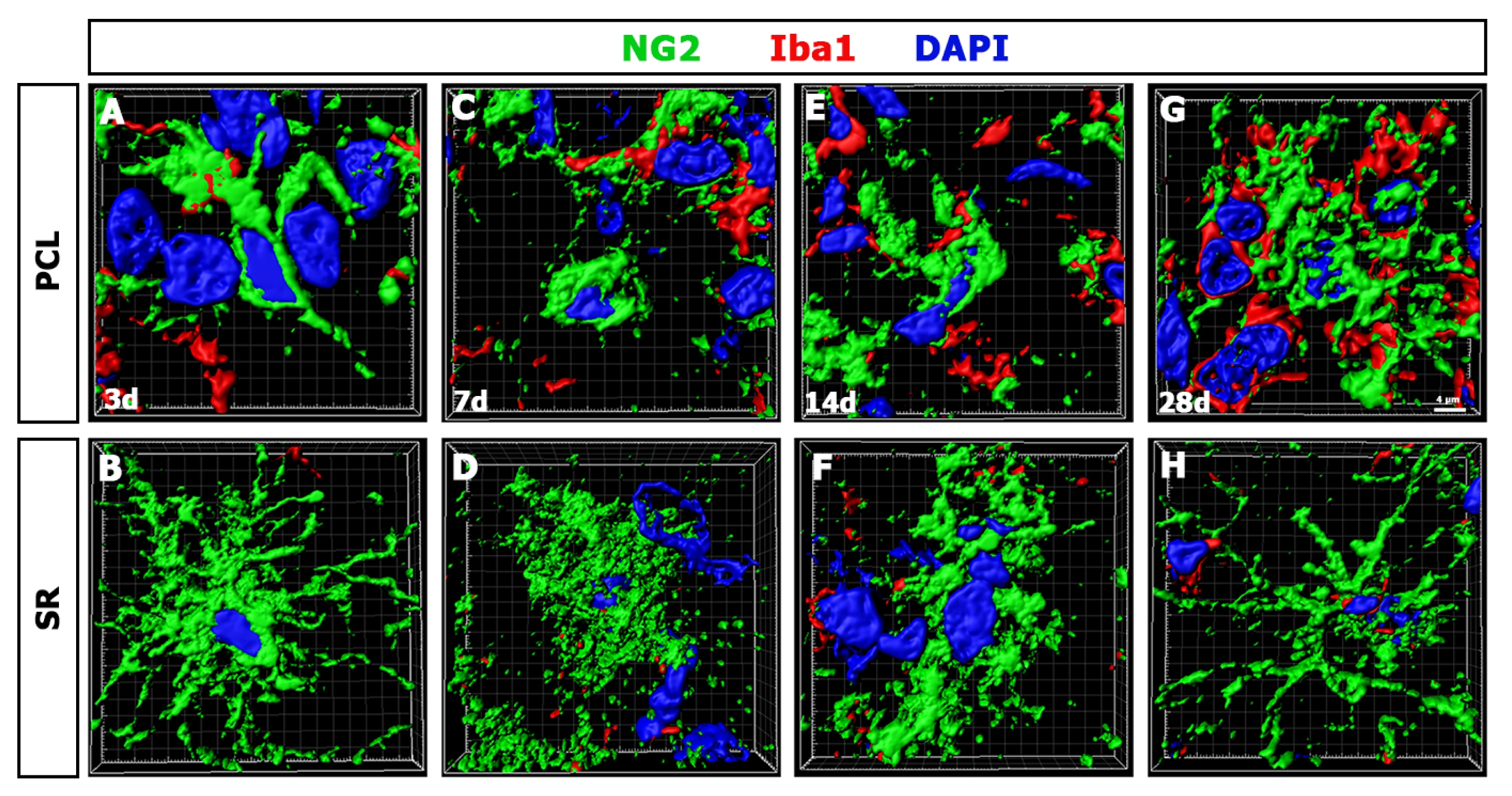
The morphological evolution of NG2 glia in the hippocampal CA1 region of rats 3, 7, 14, and 28 days following transient forebrain ischemia. Three-dimensional renderings of NG2 glia are shown in Figures 4B, C, E, F, H, I, K, and L, respectively. Note that NG2 glia exhibit obvious differences in their cellular morphology and arborization of the processes between the two layers. Additionally, note that NG2 glia in the stratum radiatum at 7~14 days after reperfusion are surrounded by amorphous clouds of NG2 staining and, thus, their individual processes cannot be discerned. Cell nuclei are stained with DAPI. Scale bars represent 4 μm in A~H.