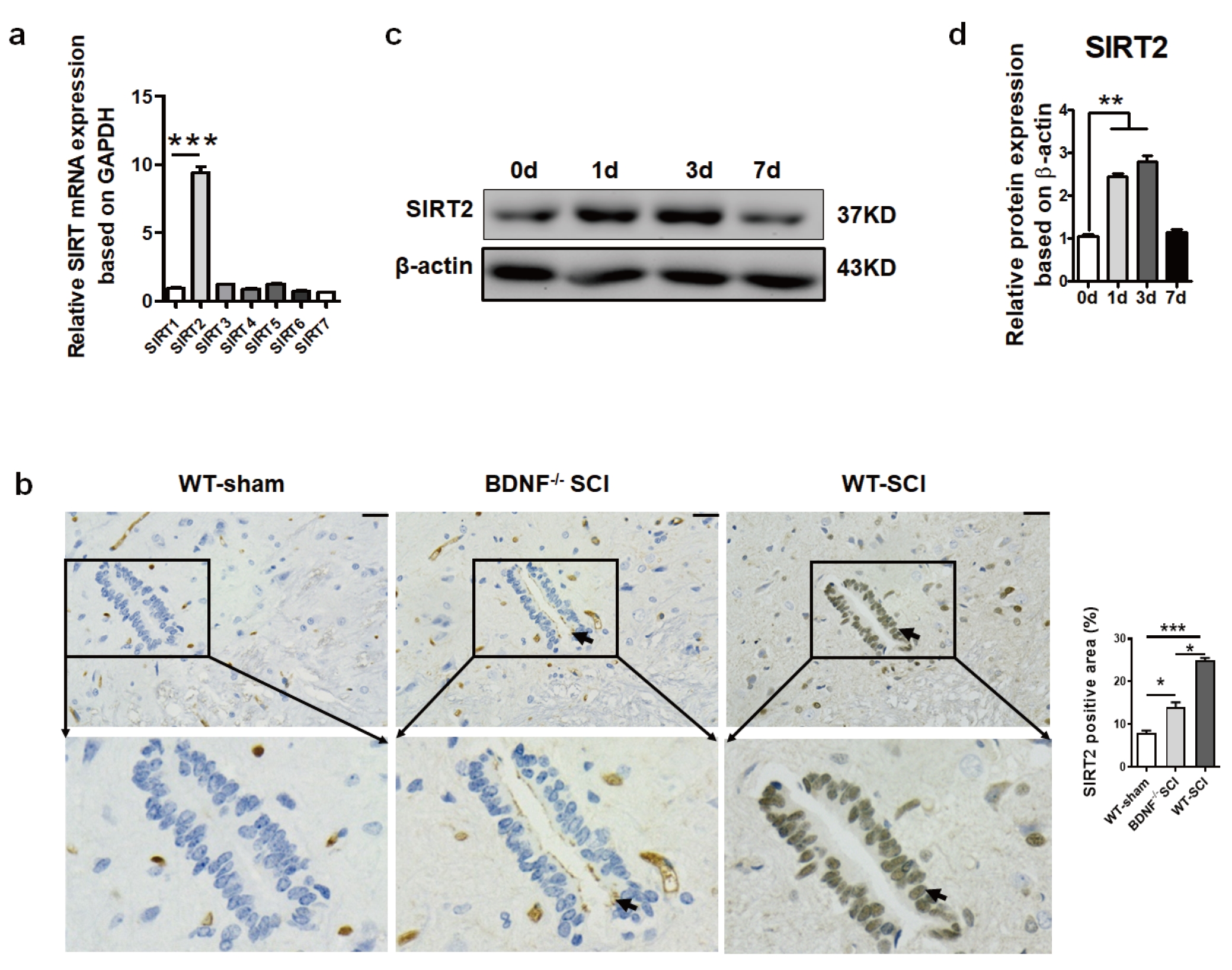
SIRT2 expression in the injured spinal cord on day 3 post SCI. (a) Spinal cord tissue was isolated from the WT mice with SCI on day 3 after SCI, and RT-PCR was performed to examine the expression of the SIRT family. SIRT2 had the highest expression level compared with other members in the spinal cord. (b) Representative IHC images of SIRT2 on day 3 after SCI in the central canal of the spinal cord (×200). Scale bars: 50 µm. The arrows indicate the positively stained cell in the central canal of the spinal cord.