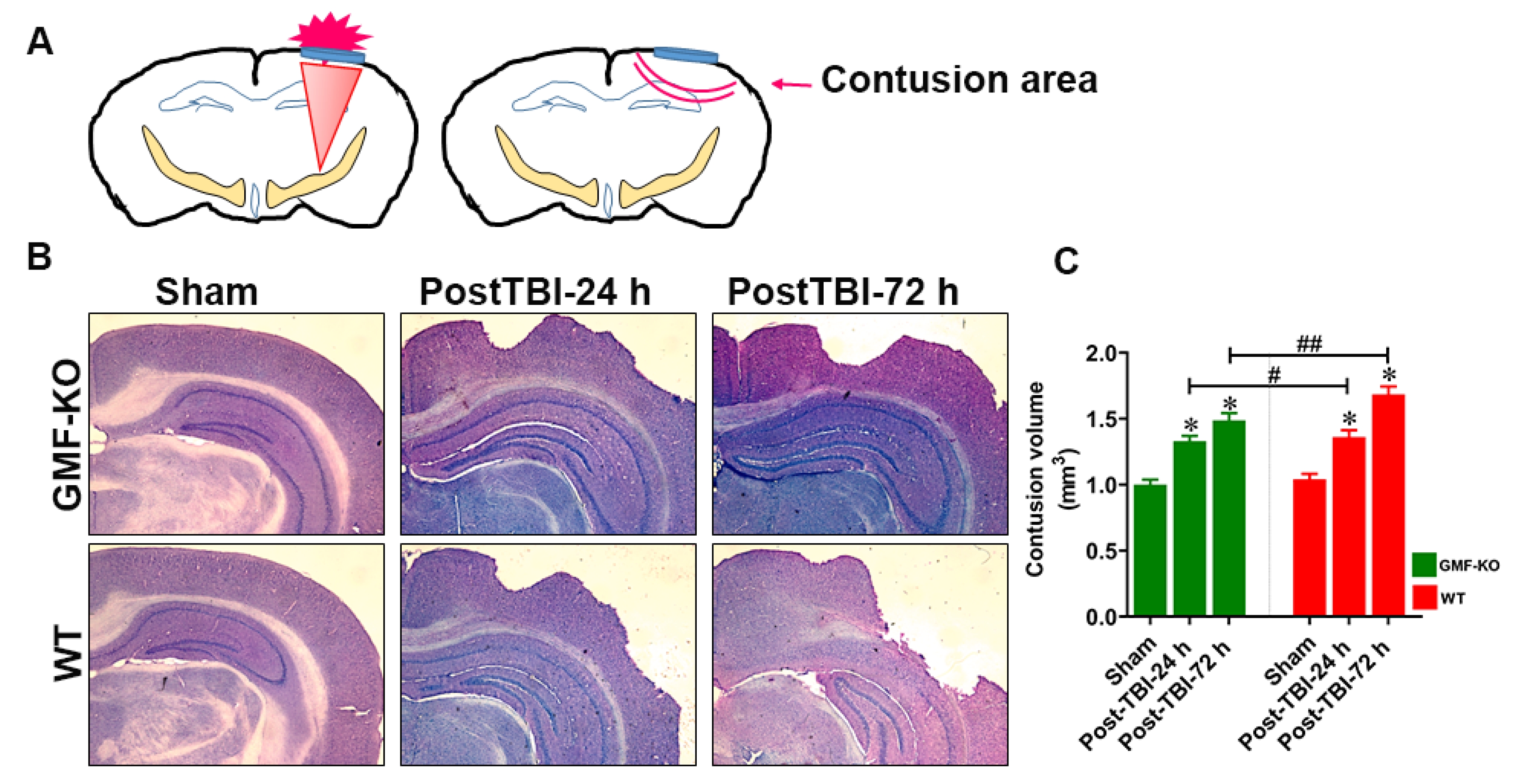
Fig. 2. Absence of GMF reduces TBI induced contusion volume and amplifies brain tissue damage. TBI caused an expanding contusion volume in WT and GMF-KO mice to brain cortical lesions. The illustration of the cortical tissue lesion after weight drop induced TBI (A). Nissl staining showed the temporal pattern of anatomical structure change (B). Bar graphs show the quantification of cortical contusion volume or cavity (C; n=6). Values are presented as mean±SEM (n=6). *p<0.05 control vs TBI subjected mice; #p<0.05 and ##p<0.01 GMF-KO TBI subjected mice vs WT TBI subjected mice.
© Exp Neurobiol