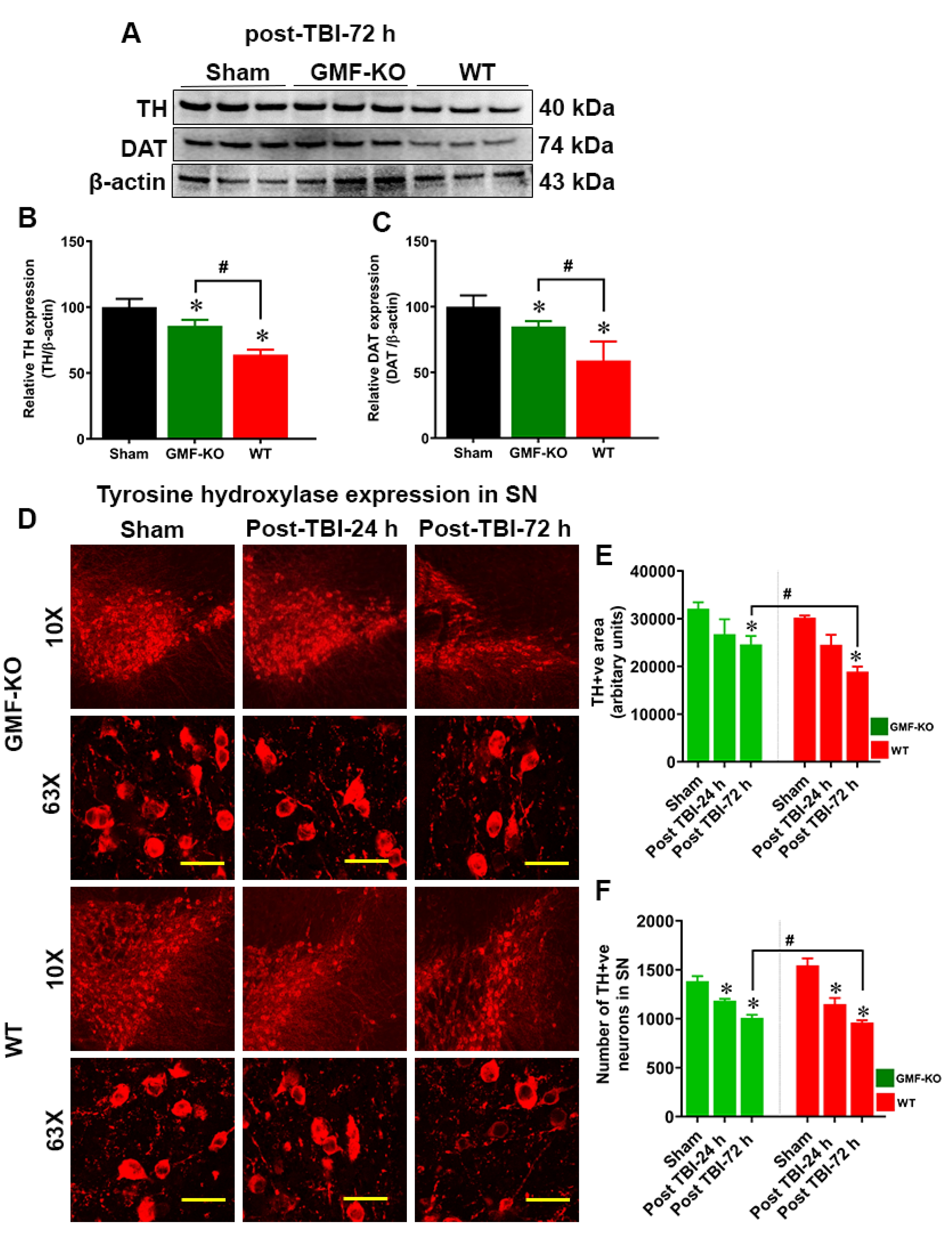
Fig. 3. Removal of GMF improves TBI induced dopaminergic markers expression in SN of the midbrain. Induction of TBI by weight drop method in the cortical region causes significant reduction in dopaminergic markers such as TH and DAT expression as determined by western blot (A) and immunofluorescence (D; red fluorescence) in the SN region of the midbrain of WT and GMF-KO mice. Representative images show the TH-positive dopaminergic neurons in the SN at different magnifications (10X and 63X). Bar graphs show the quantitation of the western blot band intensity with TH (B), DAT (C) expression, TH-positive area (E; as arbitrary units) and the number of TH-positive dopaminergic neurons (F) as compared to the controls. Values are presented as mean±SEM (n=6). *p<0.05 control vs TBI subjected mice; #p<0.05 GMF-KO TBI subjected mice vs WT TBI subjected mice.
© Exp Neurobiol