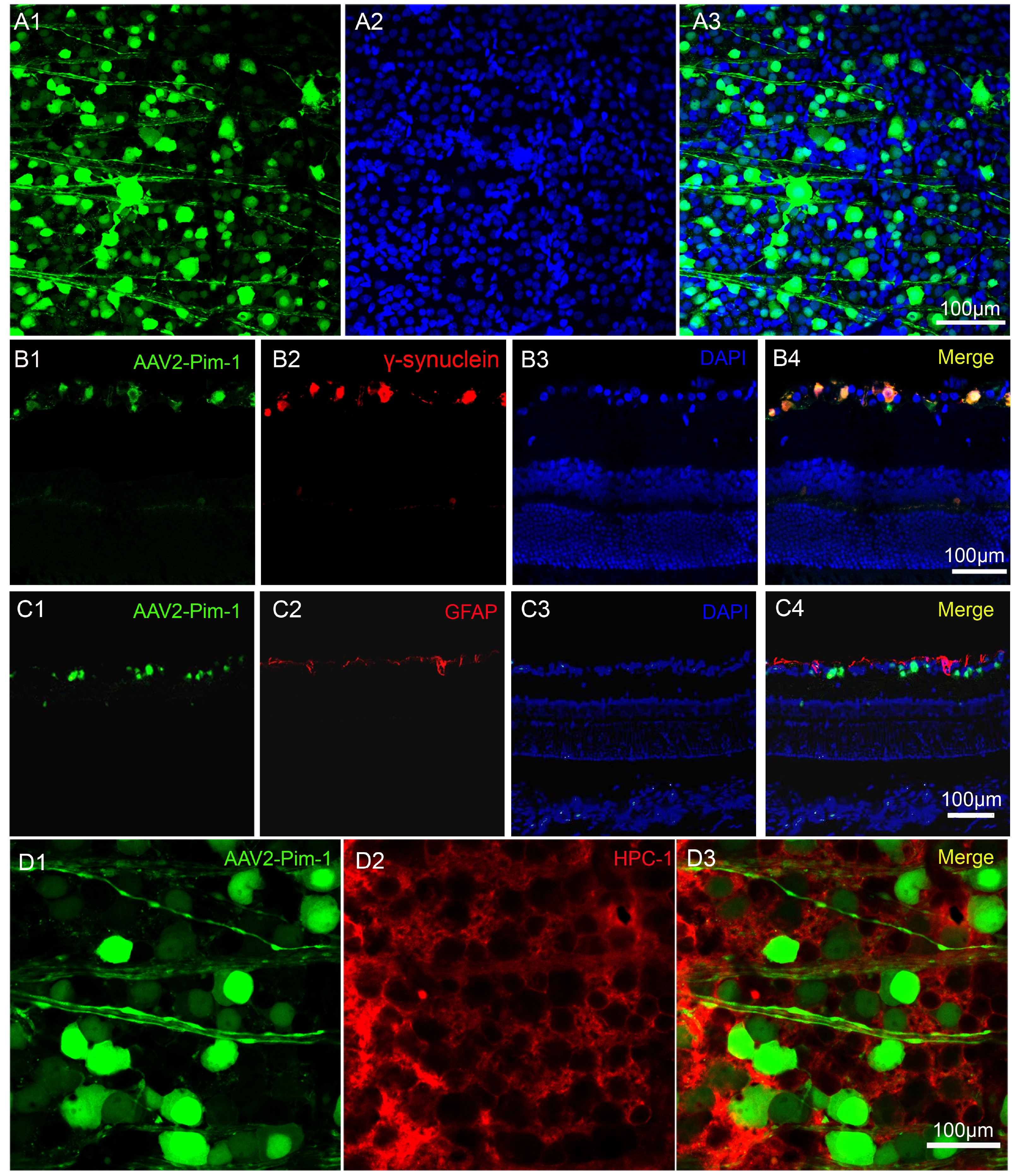
Fig. 1. Retinal flat mounts and cryostat sections of the eyeball in the AAV2-Pim-1 infected rat were stained with immunofluorescence histochemistry. (A1, B1, C1, D1) AAV2-Pim-1 infected the retina and RGCs. (A2, B3, C3) Blue staining indicates DAPI labeled nuclei. (A3, B4, C4, D3) AAV2-Pim-1 was co-located with γ-synuclein, GFAP or HPC-1. Green fluorescence for AAV2-Pim-1 and red immunostaining for γ-synuclein, GFAP or HPC-1. Scale bar=50 μm, n=4.
© Exp Neurobiol