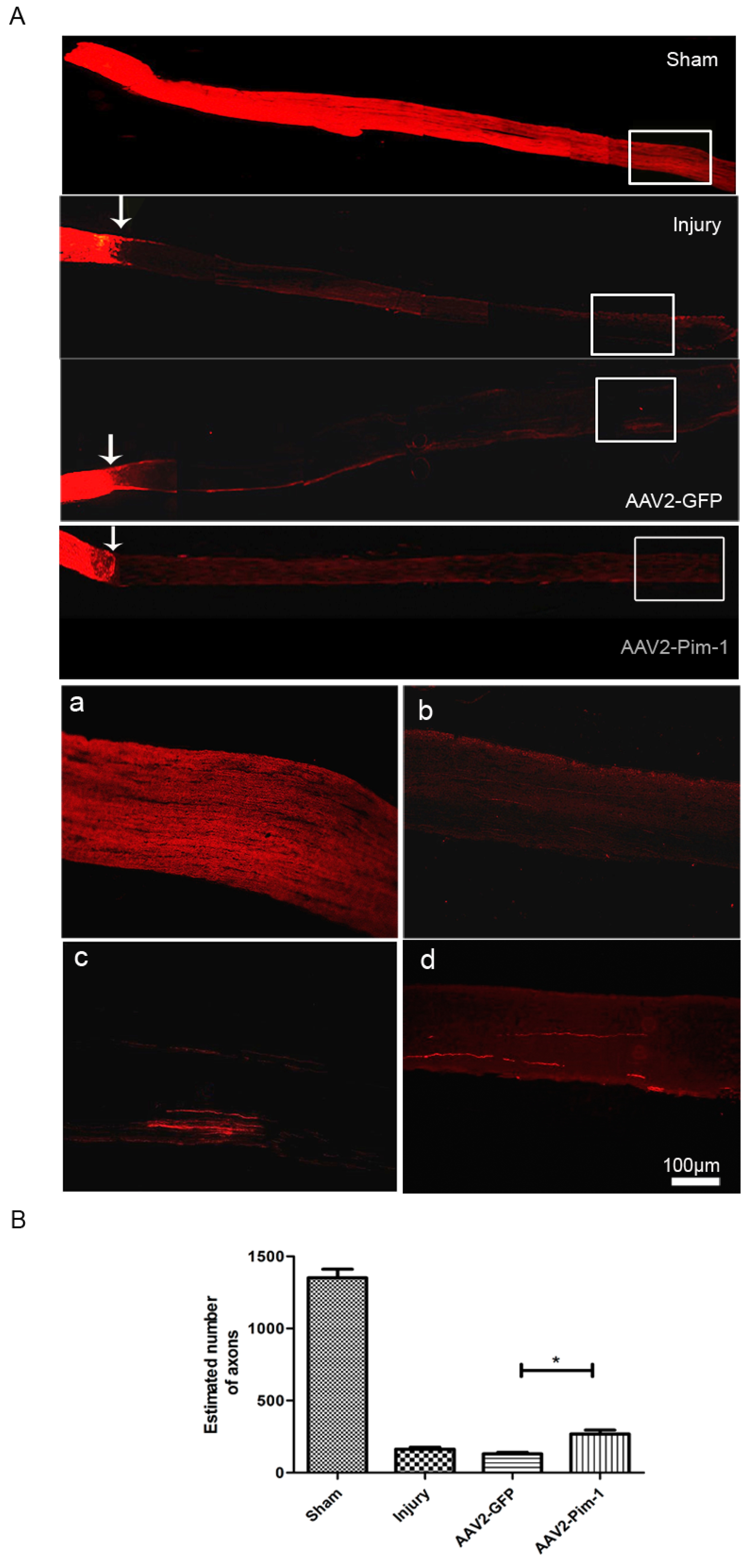
Fig. 5. Retinal ganglion cell axonal growth was stimulated by AAV2-Pim-1 in the ONC model. (A) Axons with red fluorescence intensity were clearly visible in Sham group by RITC tracing. The axons in the crush site in Injury group and AAV2-GFP group were agglutinated together (arrowhead), and a few axons were still seen in the distal segment (rectangle); meanwhile, there were more axons in the distal optic nerve passing the injured sides in AAV2-Pim-1 group. (a~d) Represent larger versions of the rectangular area of Sham, Injury, AAV2-GFP and AAV2-Pim-1 groups respectively. Scale bar=100 μm. (B) Quantitative analysis of the number of axons in the distal stump of the injured optic nerve in each group. Compared with AAV2-GFP group, *p<0.05; n=6.
© Exp Neurobiol