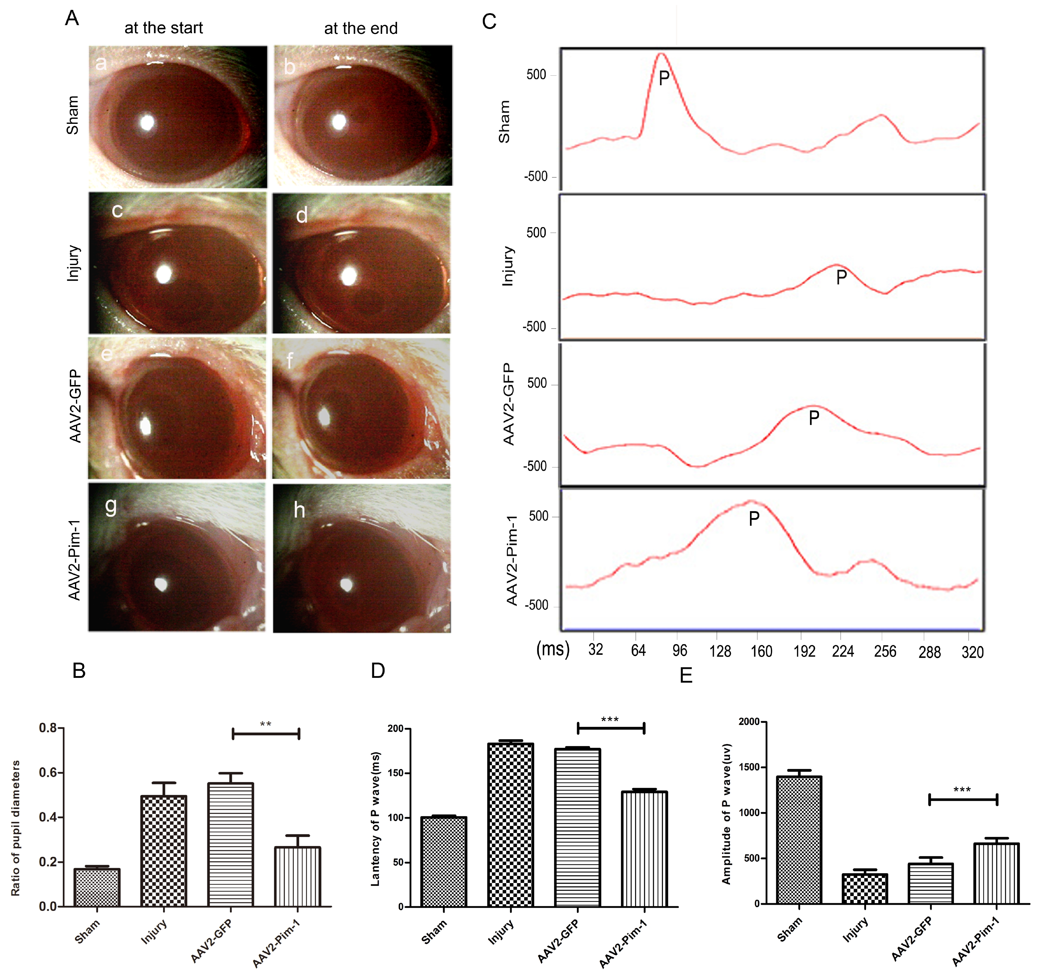
Fig. 6. The visual function is increased in vivo after intravitreal injection of AAV2-Pim-1. (A) The minimum and maximum pupil diameters were recorded in each group by pupil light reflex. Pupils were at the start (a, c, e and g) and end (b, d, f and h) of the stimulus. (B) The minimum/maximum pupil diameter ratio in Injury and AAV2-GFP was larger than that in Sham group, but the ratio in AAV2-Pim-1 group after Pim-1 overexpression was lower than that in Injury group and AAV2-GFP group. Compared with the AAV2-GFP group, **p<0.01; n=6. (C) Representative of FVEP tracings of the four groups. (D, E) The data of FVEP indicated a higher amplitude and a shorter latency in AAV2-Pim-1 group compared with AAV2-GFP group. Compared with AAV2-GFP group, ***p<0.001; n=6.
© Exp Neurobiol