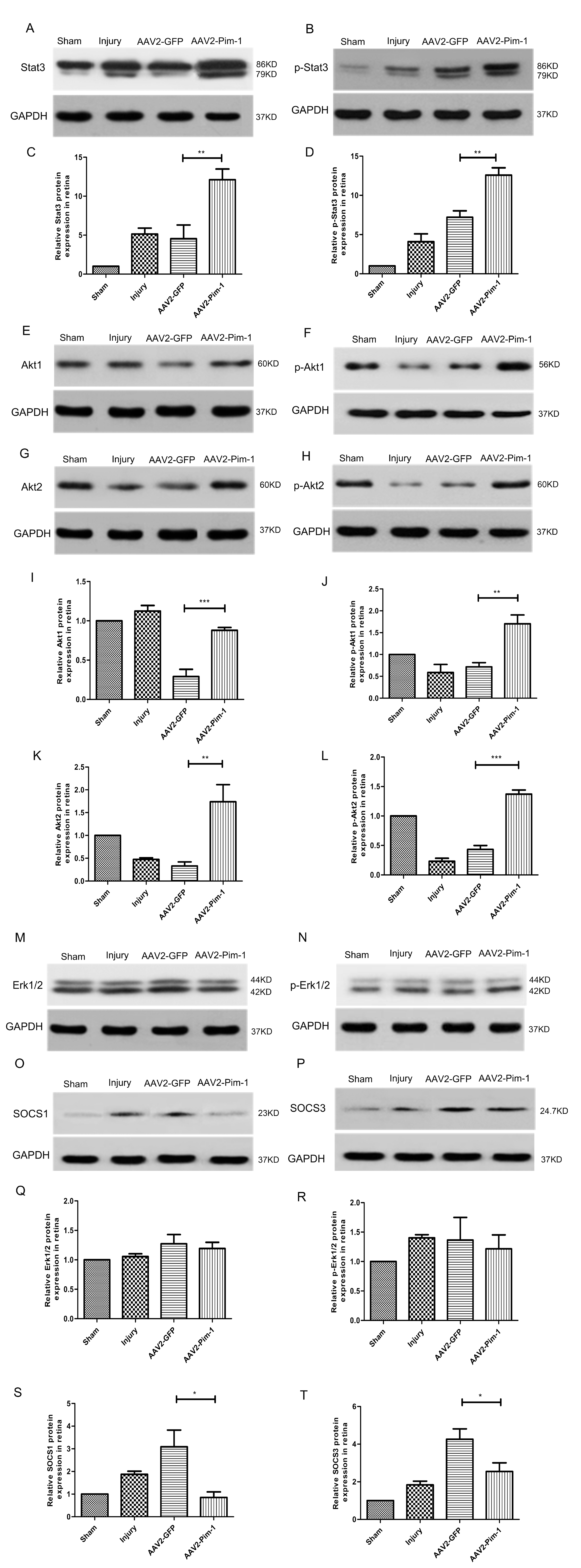
Fig. 7. Protein expressions of the key retinal signaling pathways in the ONC model in vivo. Animals were intravitreally injected with either AAV2-GFP or AAV2-Pim-1. Two weeks later, animals were subjected to ONC and retinas were harvested 2 weeks post injury. (A, B, E, F, G, H, M, N, O, P) Western blot analysis of the retinal lysates from AAV2-GFP and AAV2-Pim-1 animals using antibodies against Stat3, p-Stat3, Akt1, p-Akt1, Akt2, p-Akt2, Erk1/2, p-Erk1/2, SOCS1 and SOCS3. GAPDH served as loading control. Western blotting was used to detect the retinal protein expression. (C, D, I, J, K, L, Q, R, S, T) Quantification by densitometry of retinal lysates between the four groups for mentioned-above molecules, normalized to GAPDH loading control. Compared with AAV2-GFP group, *p<0.05, **p<0.01, ***p<0.001, #p>0.05; n=6.
© Exp Neurobiol


