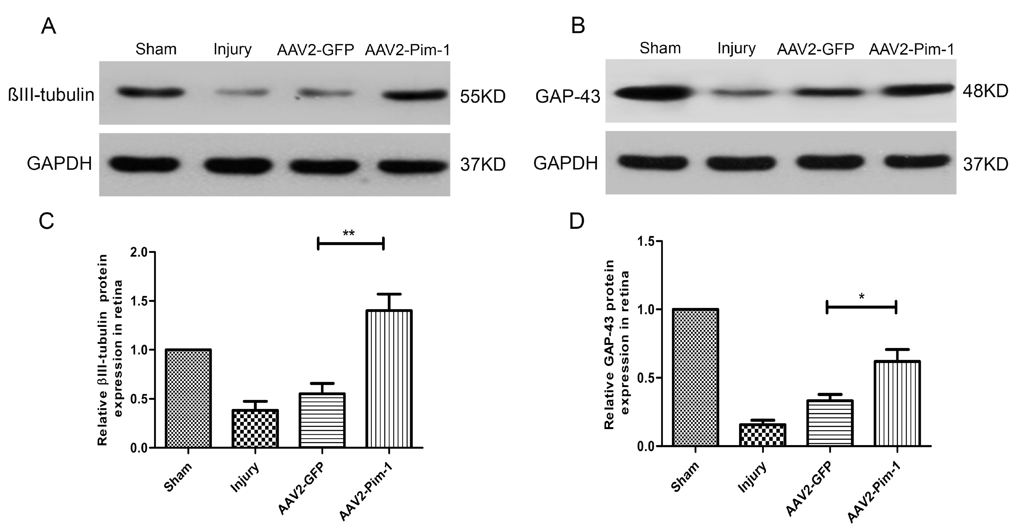
Fig. 9. Pim-1 boosted the expression of βIII-tubulin and GAP-43 axonal regeneration-related protein. (A, B) Western blot analysis of retinal lysates using antibodies against βIII-tubulin and GAP-43. (C, D) Quantification of βIII-tubulin and GAP-43 protein band intensities in the retinas treated with AAV2-Pim-1. The results were normalized to the GAPDH loading control. Compared with AAV2-GFP group, *p<0.05, **p<0.01; n=6.
© Exp Neurobiol