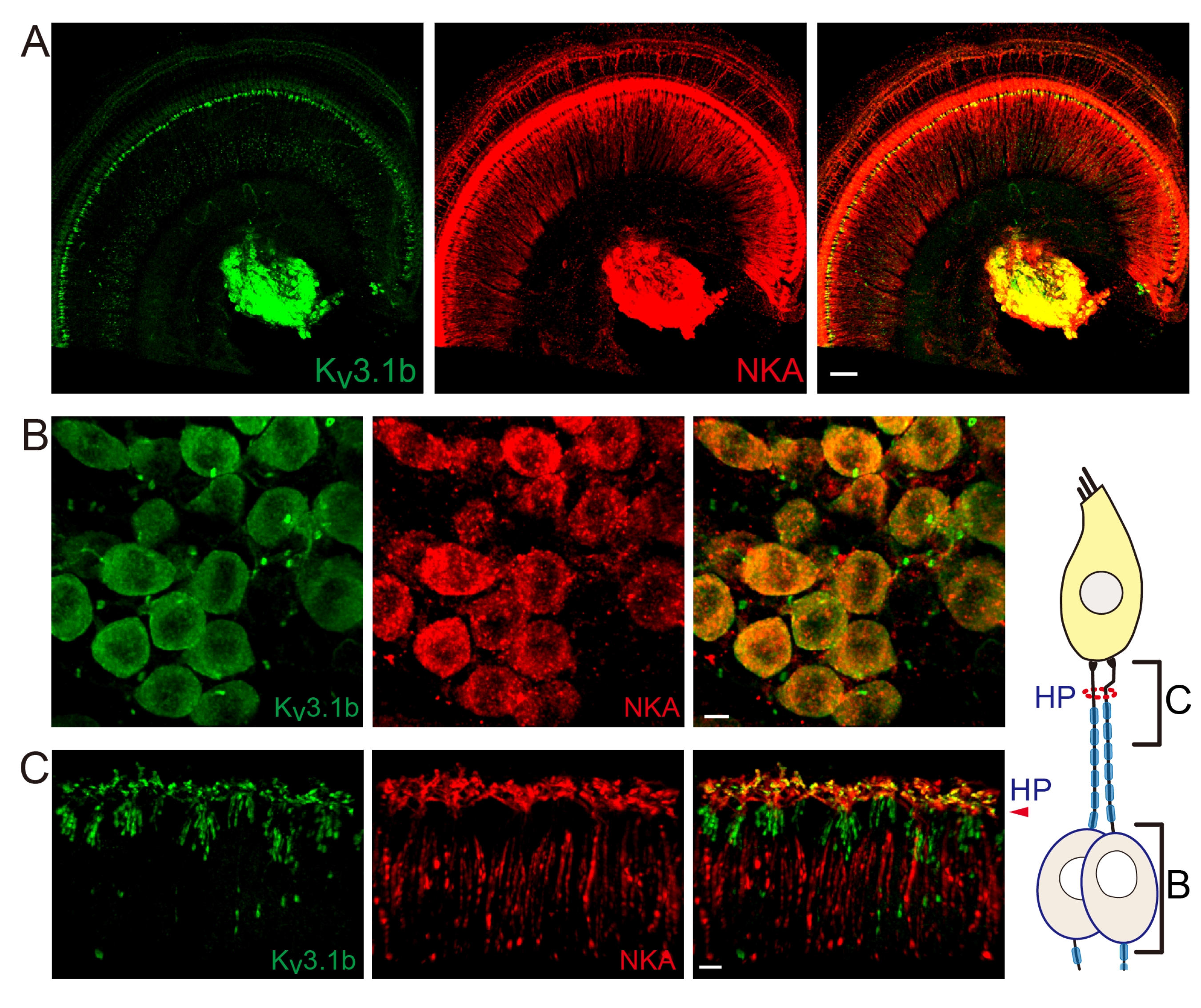
Fig. 1. Kv3.1b is expressed in the type 1 afferents. (A) Low magnification images (n=2 cochlear preparations from 2 rats) of a cochlear preparation double-immunolabeled with anti-Kv3.1b and anti-Na+, K+ ATPase α3 (NKA). Scale bar: 50 μm. (B, C) High magnification images of cell bodies (B, n=4 cochlear preparations from 3 rats) and peripheral processes (C, n=7 cochlear preparations from 4 rats) of type 1 afferents. Kv3.1b signal is present in most NKA-positive cell bodies (100%, 214 out of 214 NKA-positive SGNs) and unmyelinated dendritic segments. The relative anatomical regions corresponding to each panel B and C are described in the diagram. HP: habenula perforata. Scale bar: 5 μm.
© Exp Neurobiol