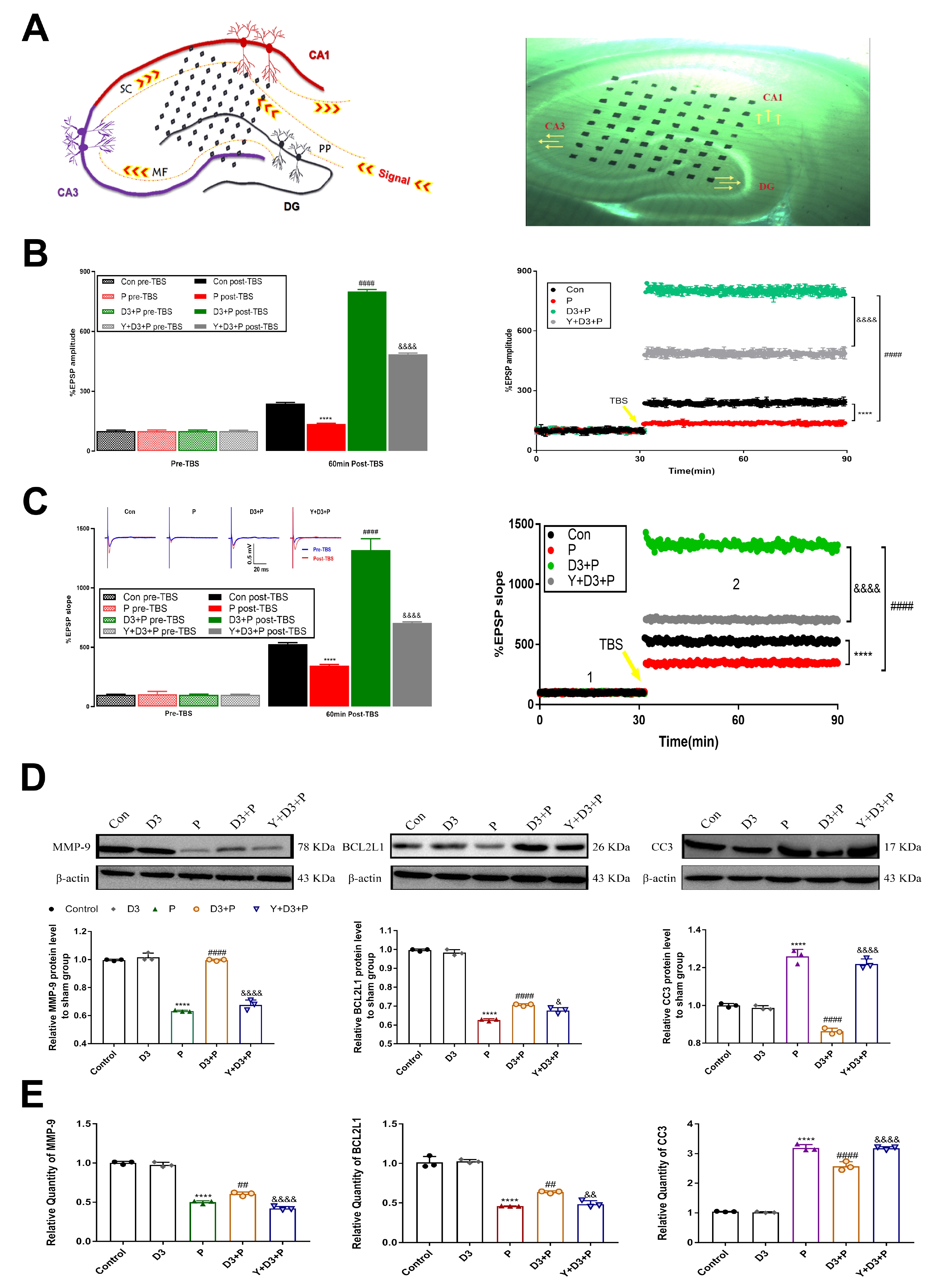
Fig. 8. Effect of a single neonatal propofol exposure on the electrophysiology and apoptotic signaling in P30 rodent hippocampi. (A) Schematic diagram, and photomicrograph of the Med 64 probe position on DG and CA1 regions. DG, dentate gyrus; MF, mossy fiber; PP, perforant pathway; SC, Schaffer collaterals. (B) The fEPSP magnitude in the ex-vivo electrophysiological assay. The long term results at P30 following a single neonatal injection of propofol or 20 μg/kg dexmedetomidine (DEX) was designated as P group or D3 group. After the co-exposure regimen executed on P7, P30 rats were designated as D3+P group (20 μg/kg DEX 1 h before propofol dosing) and Y+D3+P2 group (yohimbine 1 μg/kg 0.5 h before the similar injection regimen with DEX and propofol). Propofol significantly reduced the magnitude of the increase in LTP, indicating a deleterious effect of propofol on synaptic plasticity. DEX restored LTP in acute hippocampal slices from P30 rats. Yohimbine (Y) impaired LTP by inhibiting fEPSP, when compared to DEX-treated rats. (C) Data represent a significant increase in the fEPSP slope. (D) Western blotting of MMP-9, BCL2L1 and CC3 in the hippocampi of P30 rats. β-actin was used as internal control. (E) mRNA expression of MMP-9, BCL2L1 and CC3 in the hippocampi of P30 rats. ****p<0.0001 vs. control group, ##p<0.01, ####p<0.0001 vs. P group, &p<0.05, &&p<0.01, &&&&p<0.0001 vs. D3+P group (n=3 animals per group).
© Exp Neurobiol