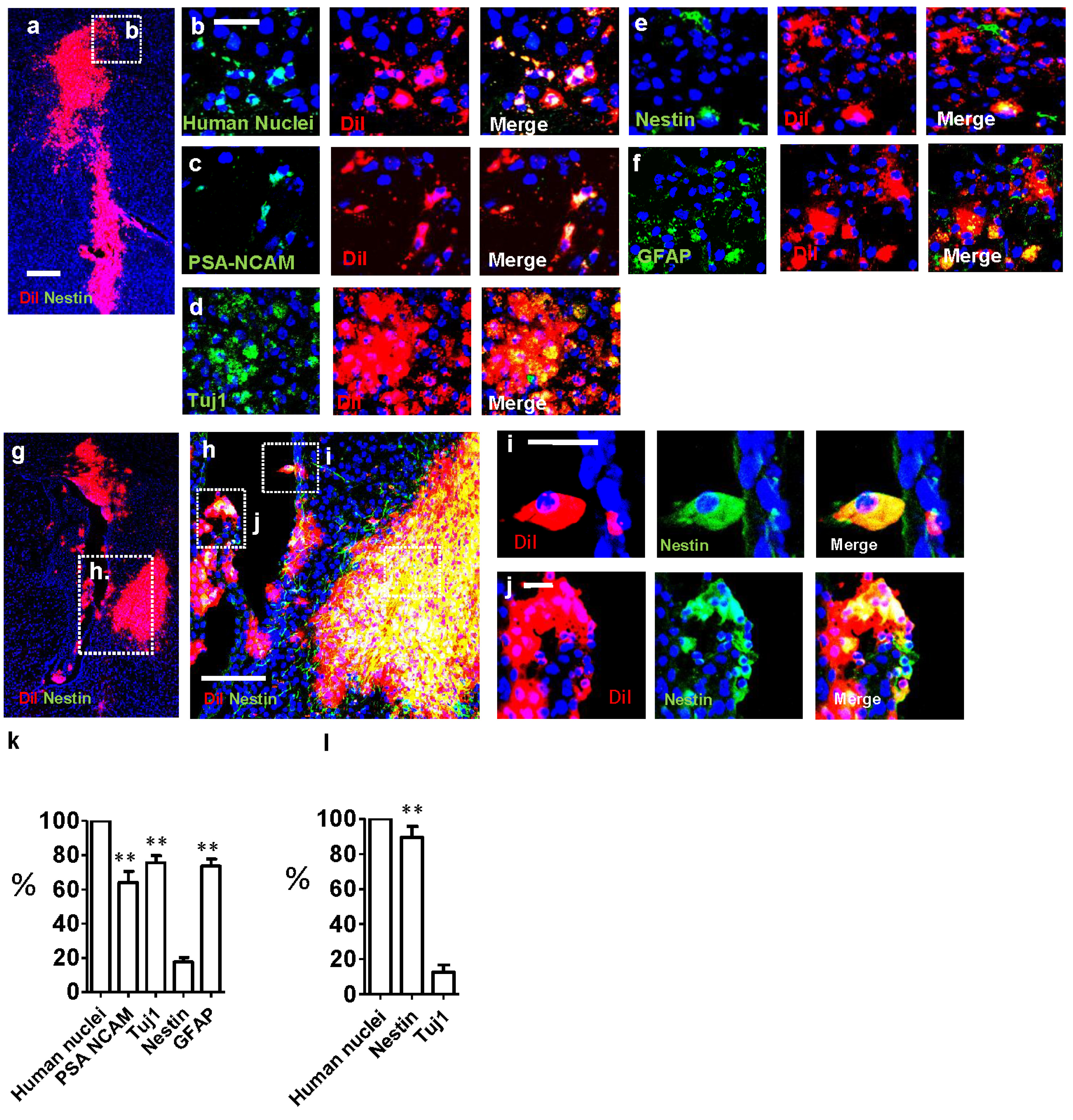
Fig. 9. Immunofluorescence analysis of CN cells engrafted into striatum of immune-deficient NOD-SCID mice. (a) Low power image of the grafted area showing DiI-labeled CN cells four weeks after engraftment. (b) Expression of hNA+ in DiI+ cells, confirming that Dil+ cells are from the CN-derived cells, not a result of contamination from the dye. (c~f) Immunofluorescence staining of CN-derived cells engrafted in the striatum near the SVZ, showing Tuj1+, PSA-NCAM+, GFAP+, and PSA-NCAM+ cells have morphology of migrating neuroblasts. (g) DiI-labeled fetal brain-derived SNSCs four weeks after engraftment in low power image. (h~i) Immunofluorescence staining of fetal brain-derived SNSCs grafted in the striatum near the SVZ shows that a majority of the cells are Nestin+. (h, l) Cellular composition of the CN-derived cells (h) and fetal brain-derived SNSCs (l), four weeks after engraftment in the striatum near the SVZ, respectively. Most of the DiI+ CN cells were PSA-NCAM+ or Tuj1+, whereas fewer cells were hNestin+ (h). Meanwhile, most of the fetal brain-derived SNSCs grafted in the striatum near SVZ were Nestin+, and very few cells were Tuj1+ (l). One-Way ANOVA analysis, ** (p<0.05) compared to Nestin (h) and Tuj1 (l). Scale bar, 50 µm (b~f) 100 µm (h) 25 µm (i, j). Three animals (N=3) were analyzed.
© Exp Neurobiol